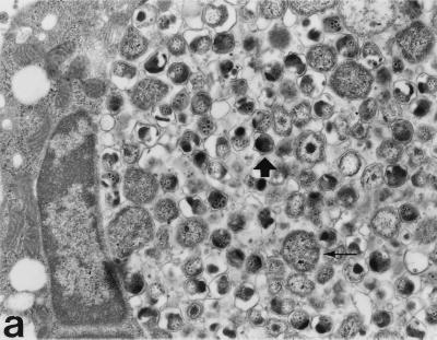
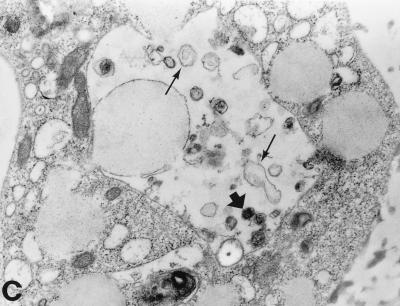
FIG. 2.
Transmission electron micrographs of C. pneumoniae-infected HL cells and monocytes 72 h postinfection. (a) Large inclusions filled with C. pneumoniae particles are found in the HL cells. Mature EBs are small, with an electrodense core (thick arrow), while RBs are large, with a coarse inner structure (thin arrow). (b) Monocytes can carry very small multiple inclusions (arrows), some containing only one C. pneumoniae particle. A thin membrane always covers these inclusions. (c) The inclusions in monocytes contain far fewer C. pneumoniae particles than do those in HL cells. Only a few mature-looking EBs can be seen (thick arrow), and the RBs are clearly transformed (thin arrows). Magnification, ×10,400.


