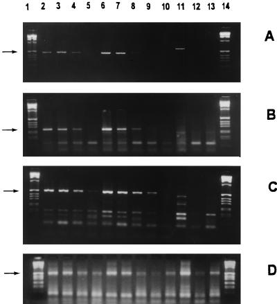
FIG. 3.
Viability of C. pneumoniae in monocytes, studied by RT-PCR. (A to C) The mRNA expression of gene transcripts for 16S rRNA (463 bp); (A) and for Crp60 (252 bp); (B) was positive up to 3 days after infection, and that for HSP60 (507 bp); (C) was positive up to 7 days after infection. (D) The chromosomal β-actin gene of monocytes was expressed at all the time points. IFN-γ treatment of the infected monocytes seemed not to be able to inhibit gene expression. The arrows indicate the amplification products. Lanes: 1 and 14, molecular weight markers; 2 to 5, infected monocytes; 6 to 9, infected, IFN-γ treated cells; 10 to 13, uninfected cells. For each set of lanes, the samples were obtained right after infection and on days 1, 3, and 7 after infection, respectively.

