FIG. 5.
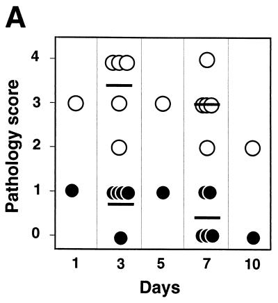
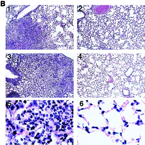
Mouse lung pathology induced by wt and ΔcyaA B. bronchiseptica. BALB/c mice were inoculated in parallel with those for Fig. 3. Histological sections of lung tissues were prepared as described in Materials and Methods. (A) Samples were submitted for observation in a blinded fashion. Upon examination, samples were given scores of 0 (no pathology), 1 (mild inflammation in ≤10% of the bronchioles and/or ≤10% of lung tissue), 2 (inflammation in 10 to 30% of bronchioles and/or mild inflammation in 10 to 30% of lung tissue), 3 (inflammation in >30% of bronchioles and mild to moderate inflammation in >30% of lung tissue), and 4 (inflammation in >50% of bronchioles and moderate to severe inflammation in >30% of lung tissue). Bars indicate the averages of five animals. Open circles represent animals infected with RB50 (wt), and solid circles represent animals infected with RB58 (ΔcyaA). (B) Representative sections of lungs infected with wt (panels 1 and 3) or ΔcyaA (panels 2 and 4) bacteria on day 3 (panels 1 and 2) or day 7 (panels 3 and 4). Magnification, ×100 (panels 1 to 4) and ×1,000 (panels 5 and 6).


