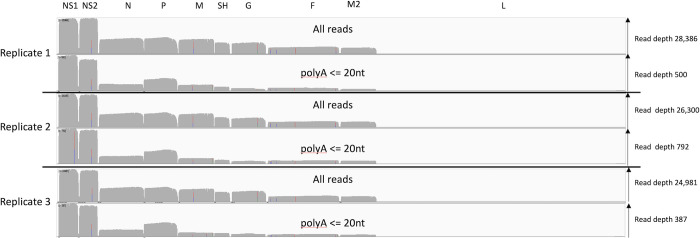
Fig 6. Calu-3 infected cell transcripts mapped to the RSV genome and grouped by poly A tail length.
An IGV generated image of the dRNAseq reads mapped to the viral genome split into three sections representing the mRNA sequenced from Calu-3 infected cell replicates 1, 2 and 3, the location of the viral ORFs is indicated along the top. Each section has two panels, the top panel shows the overall depth of reads along the genome for all the mapped reads and the lower one for only reads with a poly A length of between 1 and 20 nucleotides as reported by nanopolish software.

