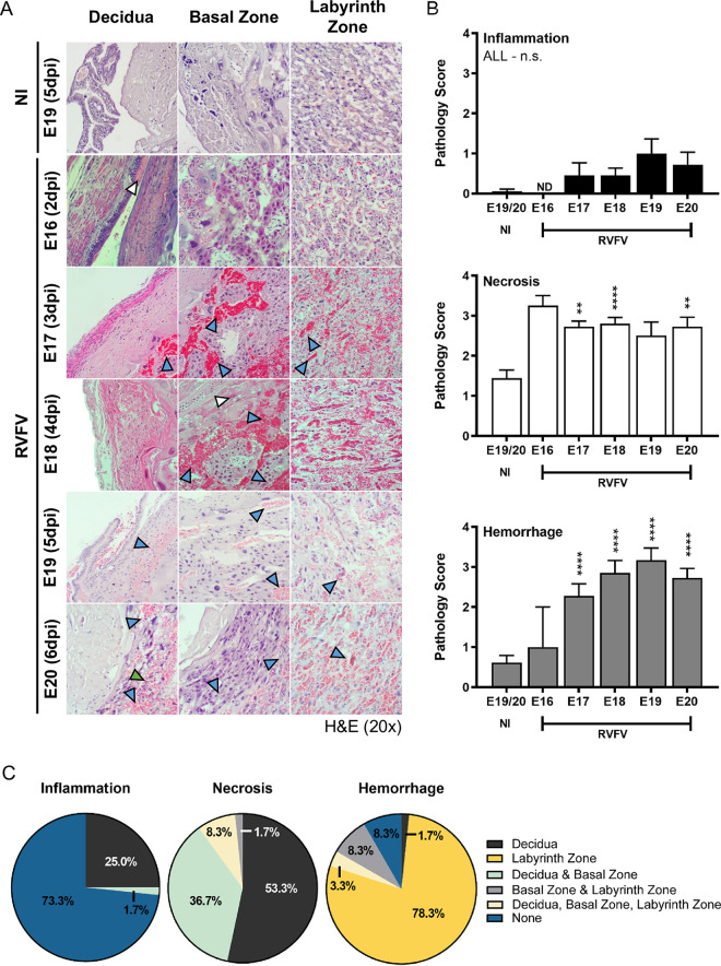
Fig 4. RVFV-mediated pathology in the placenta of dams that succumb to infection as early as E16 (2dpi).
(A) Pathology of infected placentas (n = 2, 11, 20, 6, 11, respectively) from dams succumbing to disease from E16-E20 (2-6dpi) based on H&E staining. N = 18 placentas were collected for uninfected (NI) controls. Blue, white, and green arrow heads indicate hemorrhaging, necrosis, or leukocyte inflammation, respectively. (B) Pathology scores (scale: 0–4) of inflammation (top), necrosis (middle), and hemorrhaging (bottom). (C) Pie graph representing the percentage of placentas from all gestations (E16-22) with evidence of inflammation, necrosis, or hemorrhage in the decidua, basal zone, labyrinth zone, or multiple layers from RVFV infected dams. NI = no infection. ** = p < 0.01, **** = p < 0.0001, & n.s. = not significant. ND = none detected. In (B), an ANOVA with multiple comparisons was performed to determine statistical significance between the uninfected cohort and infected groups at each time point.

