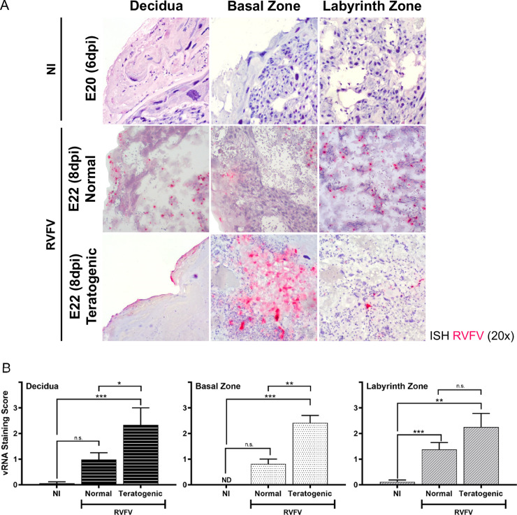
Fig 5. Teratogenicity is associated with widespread infection of multiple placenta layers.
(A) ISH (magenta) stained placentas (E20 & E22) from uninfected (NI) and infected dams. Infected placentas are segregated into those from pups with normal appearance (herein “normal”) or those from pups with gross signs of teratogenicity (herein “teratogenic”). Panels are separated based on placenta regions: decidua, basal zone, and labyrinth zone. Tissues were hematoxylin counterstained. 20x images. (B) Severity of ISH staining (scale: 0–3) in normal and teratogenic pups based on individual regions of placentas collected between E19-22. * = p < 0.05, ** = p < 0.01, *** = p < 0.001, & n.s. = not significant. ND = none detected. An ANOVA with multiple comparisons was performed to determine statistical significance between groups. NI (n = 6), normal (n = 15), teratogenic (n = 4).

