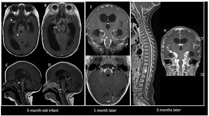
Figure 1. Neurocutaneous melanosis. A 5-month-old infant, without pigmented skin lesions, who was referred due to acute hydrocephalus. Axial (A, B) and sagittal (C, D) pre and post-contrast T1WI demonstrated subtle foci of spontaneous hypersignal in the temporal uncus, brainstem and cerebellar vermis (thin white arrows), with extensive leptomeningeal enhancement (↦). In a control one month later, progression of leptomeningeal enhancement was observed, which was predominantly infratentorial (arrowheads in E, F). A control three months later (G, H) showed aggressive spinal and encephalic progression of leptomeningeal dissemination (thick white arrows).

