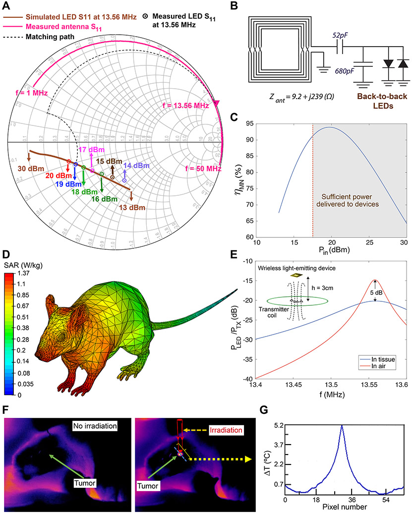
Fig. 3. Tuning wireless power transfer efficiency and evaluating safety for photothermal therapy in freely-behaving mice.
(A) Smith chart showing simulated and measured S11 for back-to-back devices with LEDs emitting 810 nm wavelength light. The measured S11 of the antenna is also shown along with the ideal matching path (dashed line). (B) L-match structure and matching components for the back-to-back devices. The 52 pF capacitance was constructed by placing two 22 pF and 30 pF capacitors in parallel. (C) Variation of the matching network efficiency, ηMN, with input power (Pin), assuming capacitor quality factor of Qc=100 for the matching components. (D) Quantitative evaluation of mouse body interactions with 13.56 MHz RF waves to evaluate safety of the wireless power transfer in our approach during wireless therapy cycles (compare with SAR values calculated for conventional 915 MHz RF waves in Supplementary Fig. 20). The scale bar is logarithmic. (E) Effect of the tissue on the wireless link efficiency (ratio of the power received at the input of the devices to the transmitted power) after implantation on the mouse skull, suggesting ~ 5 dB degradation in the power delivered to the RX antenna, due to the tissue interaction. (F) In vivo open-skull thermal images of the brain tumor, 24 h after intratumoral injection of nanoparticles. NIR emitting device was fixed above the brain (also see Supplementary Figs. 22-23). (G) Temperature variation along the blue dotted line on the brain shown in (F), verifying that only the tumor area which contained the nanoparticles was heated during the NIR irradiation (~30 mW), due to the nanoparticles photothermal response. We did not observe any elevated temperature in surrounding normal brain tissues.

