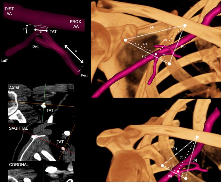
Fig. 1.
Measurement methodology presented in graphical form. To determine the TAT morphology, the multiplanar reconstruction of CT images was performed. PROX AA Proximal axillary artery; DIST AA distal axillary artery; TAT thoracoacromial trunk; Delt deltoid branch of TAT; LatT lateral thoracic artery; Pect pectoral branch of TAT; (1) Length of TAT; (2) maximal diameter of TAT; (3) TATs’ ostial area; (4) distance from the coracoid process to the clavicle, (5) distance from the coracoid process to the second rib, (6) distance from the clavicle process to the second rib, (7) distance from the coracoid process to the TAT, (8) distance from the clavicle to the TAT, and (9) distance from the second rib to the TAT. An asterisk (*) indicates the length of the corresponding consecutive Pect of TAT or its branches

