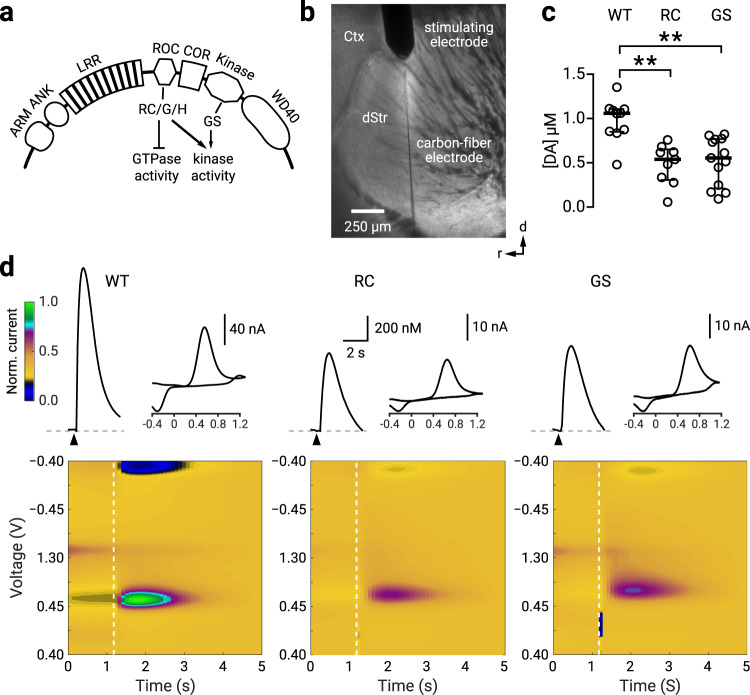
Fig. 1. RC and GS LRRK2 mice have decreased nigrostriatal dopamine release.
a A schematic diagram of LRRK2 protein: a family of Ras-like G-proteins with functionally distinct multi-domains, consisting of armadillo repeats (ARM), ankyrin repeats (ANK), the leucine-rich repeats (LRR), the C-terminal of Roc (COR), the ROC GTPase domain where the R1441C/G/H (RC) mutations reside, and the kinase domain where the G2019S (GS) mutation resides. The WD40 domain is involved in membrane binding with ARM and ANK believed to stabilize the electrostatic surfaces of the domains. b. Brightfield photomicrograph of a parasagittal slice with a concentric stimulating electrode placed in the dorsal striatum (dStr); a carbon fiber electrode was inserted adjacent to the stimulation site. Ctx, cortex. c Population data showing for evoked [DA] in WT, RC, and GS LRRK2 KI mice. The median evoked concentrations were: [DA]WT = 1057.6 ± 117.9 nM (n = 10); [DA]RC = 538.7 ± 143.2 nM (n = 9); [DA]GS = 554.9 ± 218.4 nM (n = 13). Compared to WT mice, evoked dopamine [DA] was decreased in RC mice (p = 0.006, Mann–Whitney U test). Similarly, compared to WT mice, [DA] was less in GS mice (p = 0.001; Mann–Whitney U test). There was no difference in evoked release between RC and GS mice (p = 0.69; Mann–Whitney U test). d Representative time courses for [DA] with corresponding cyclic voltammograms and color maps of fast-scanning cyclic voltammetry recordings. Peak evoked responses: [DA]WT = 1223.7 nM; [DA]RC = 655.1 nM; [DA]GS = 710.4 nM. Colormaps are normalized to the oxidation current from the WT recording. ** denotes p < 0.01. See Supplementary Table 1 for complete sample sizes by sex and statistical results.

