Figure 4.
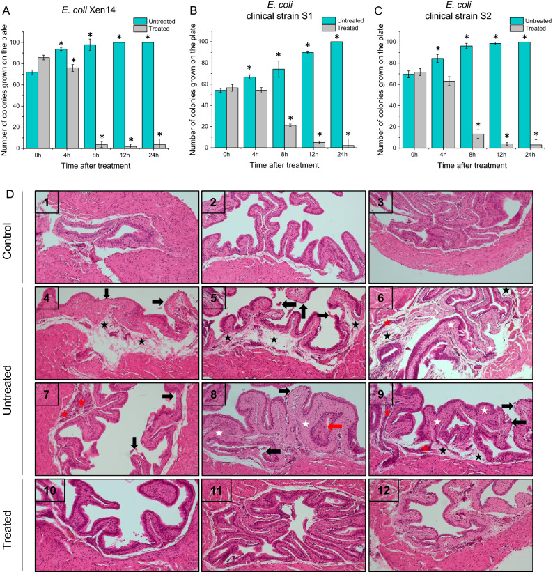
Decrease in E. coli Xen14 colonies in urine culture from mice post CSA-13 IRDye800CW treatment (grey columns) when compared to urinary tract infected untreated mice (green columns) (A) and decrease in E. coli clinical strains (S1, (B) and S2 (C), colonies in urine culture from mice post CSA-13 treatment (grey columns) when compared to urinary tract infected untreated mice (green columns). Histological analysis of mice bladder tissues: (1–3) normal murine bladder; (4–9) murine bladder infected with E. coli Xen14; tissue edema (black star), exfoliation of transitional epithelial cells (black arrow), invasion of inflammatory cells in the mucosa (red star), and bladder mucosa hyperplasia (red arrows) (10–12) Murine bladder infected with E. coli Xen14 and treated with CSA-13 (D).

