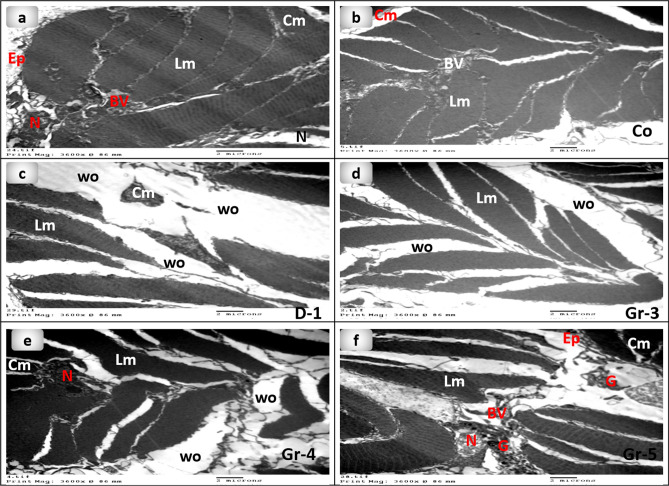
Figure 9.
Transmission electron microscopy micrographs of earthworms (Lumbricus castaneus) (a) normal earthworm; (b) worms received Vaseline, showing fissure (raw); (c) worm on the first day of injured showing the coelomic fluid emerged as well as the blood surrounds the wound appearance (circle), and (d) worms received 5 mg, (e) worms received 10 mg showing fissure(raw), (f) worms received 15 mg. Cm Circular muscle, Ep Epidermis, BV Blood vessel, G Granules, Lm Longitudinal muscle, N Nucleus, Wo Wound.

