
Figure 5. Preparation of electrohydrodynamic (EHD)-jetting and characterization of AMD3100-SPNPs.
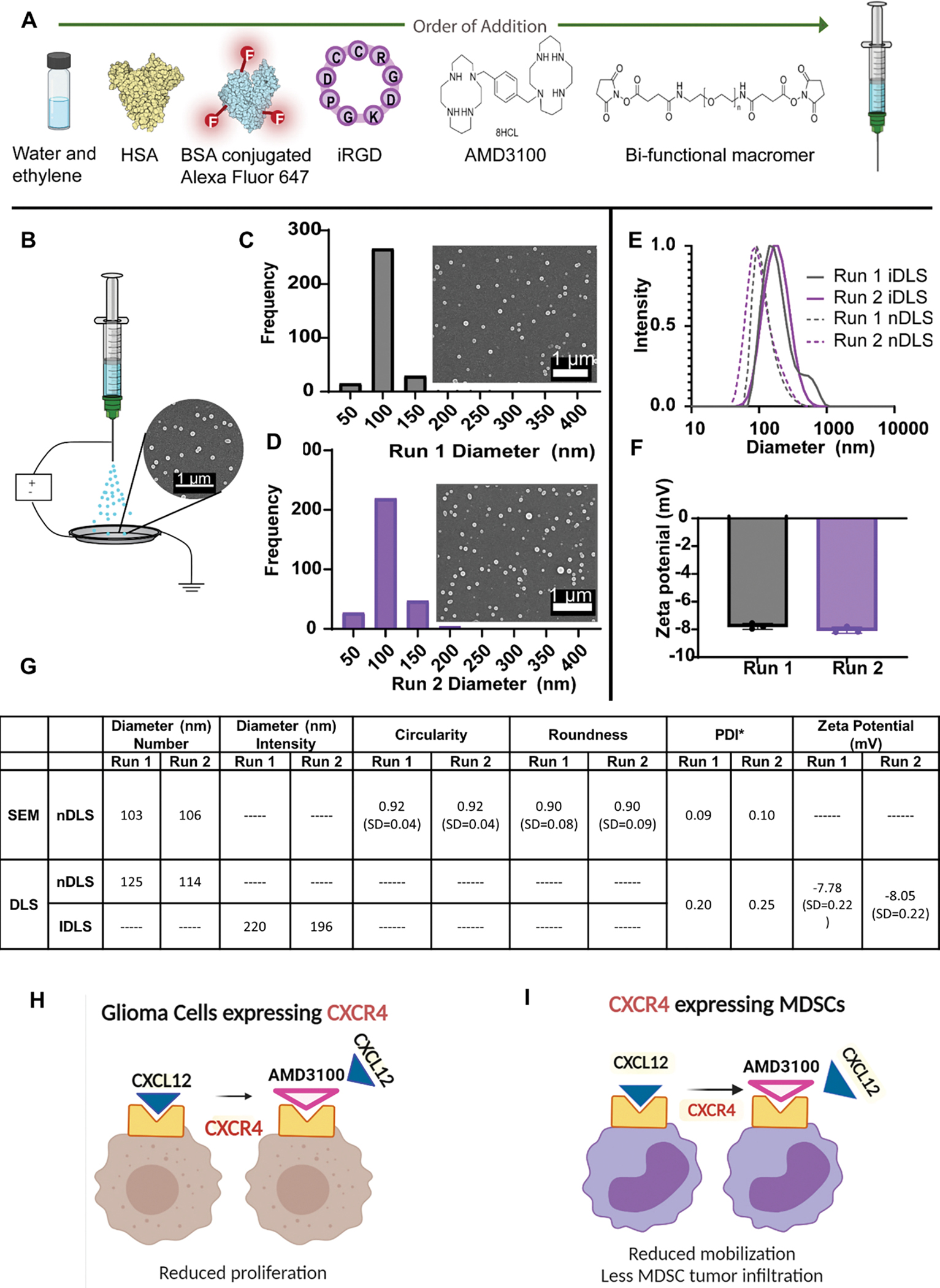
(A) Formulation of AMD3100-SPNPs indicating the order of addition of different components. (B) Schematic of the jetting process for AMD3100-SPNPs depicting a Scanning Electron Microscopy (SEM) image of the SPNPs jetted atop of the collection plate (scale bar = 1μm). (C) Size distribution of SPNPs of an independent run, Run 1, in their dry state characterized via SEM and ImageJ analysis. Average diameter, 103 ± 20 nm (PDI = 0.09). Scale bar = 1 μm. (D) Size distribution of SPNPs of a second independent run, Run 2, in their dry state characterized via SEM and ImageJ analysis. Average diameter, 106 ± 25 nm (PDI = 0.10). (E) Numbers based Dynamic Light Scattering (nDLS) size distribution (dashed) and Intensity based DLS (IDLS) of SPNPs in PBS comparing Run 1 and Run 2. (F) Zeta potential of Run 1 and Run 2. (G) Summary table of SPNP characterization of size, shape and charge. (H, I) Schematics represent the therapeutic advantages of blocking CXCR4 in glioma cells. (H) and myeloid cells (I).