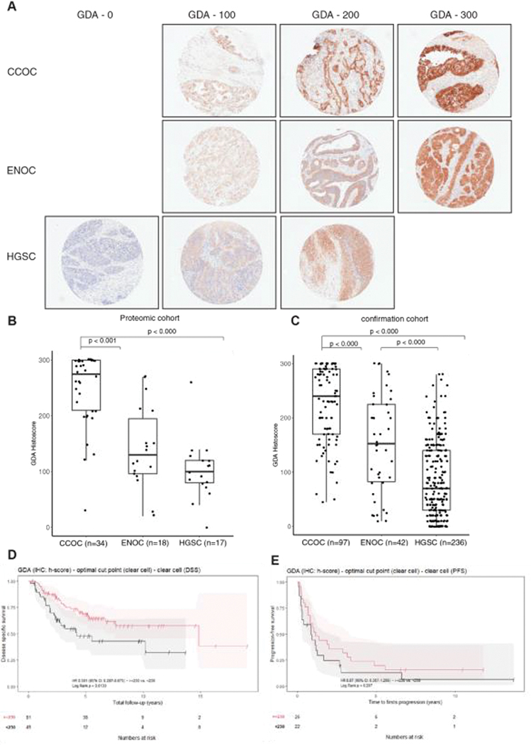
Figure 4.
Guanine deaminase (GDA) immunohistochemical validation. (A) Representative immunohistochemistry staining of GDA in different ovarian cancer subtypes, and boxplots representing (B) GDA histoscores in proteomic cases, and (C) an independent confirmation cohort. (D) Disease specific survival and (E), progression-free survival in CCOC with high GDA and low GDA expression. The statistical significance in multiple group comparisons is calculated with a Kruskal-Wallis test with a post-hoc Dunn’s test with Benjamini-Hochberg correction.

