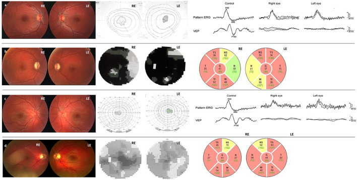
Figure 1.
(a) Case 1 Fundi at the disease onset: hyperemic optic disks, without vessel tortuosity, and with intraretinal hemorrhage at the superior arcade, central scotoma in the visual field, and normal PERG, delayed and decreased VEP 100. (b) Case 1 at the last check-up 14 years after the disease onset: pale and atrophic optic disks, small fenestrations in the central scotoma in the visual field, and thinning of the peripapillary RNFL. (c) Case 2 Fundi at the disease onset: Hyperemic optic disks, tortuotic blood vessels, central scotoma in the visual field, reduced PERG N95, delayed and decreased VEP 100. (d) Case 2 at the last check-up 12 years after the disease onset: Pale and atrophic optic disks, decreased visual field scotoma, and thinning of the peripapillary RNFL despite significant VA improvement.

