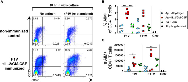
Figure 3.
Plague antigen F1V-specific CD4+ T cells as detected by activation-induced marker (AIM) assay. Cells from the draining lymph nodes 7 days post-immunization from immunized mice were cultured for 18 hours in the presence or absence of recombinant F1V antigen. Cells were then stained for surface markers and evaluated for antigen-induced upregulation of both OX40 and CD25. The frequency of specific AIM+ cells (% AIM+ of CD4+ T cells shown in Figure 2B) was calculated as the frequency of CD4+B220- T cells that specifically express both OX40 and CD25 (OX40+CD25+ double-positive population in upper right quadrant) cells following 18 hours of culture in the presence of F1V antigen minus the background frequency of OX40+CD25+ cells from the same sample following 18 hour culture in the absence of antigen. (A) Representative flow cytometry analysis of OX40 and CD25 expression by CD4+ gated T cells, of a control mouse (injected with alhydrogel but not immunized with antigen) and a F1V + IL-2/GM-CSF cytokine immunized mouse. (B) Chart shows the percent AIM+ of CD4+ T cells in the dLNs, which is the frequency of cells that are OX40+CD25+ cells in the F1V-restimulated minus the no-antigen culture for each individual sample. (C) Chart shows the number of AIM+ F1V-specific OX40+CD25+ CD4+ T cells in the dLN. n=5 per group per experiment at each time point. Statistically significant p values were determined using a two-tailed unpaired Student t test; *p < 0.05, **p < 0.01, ***p < 0.001.

