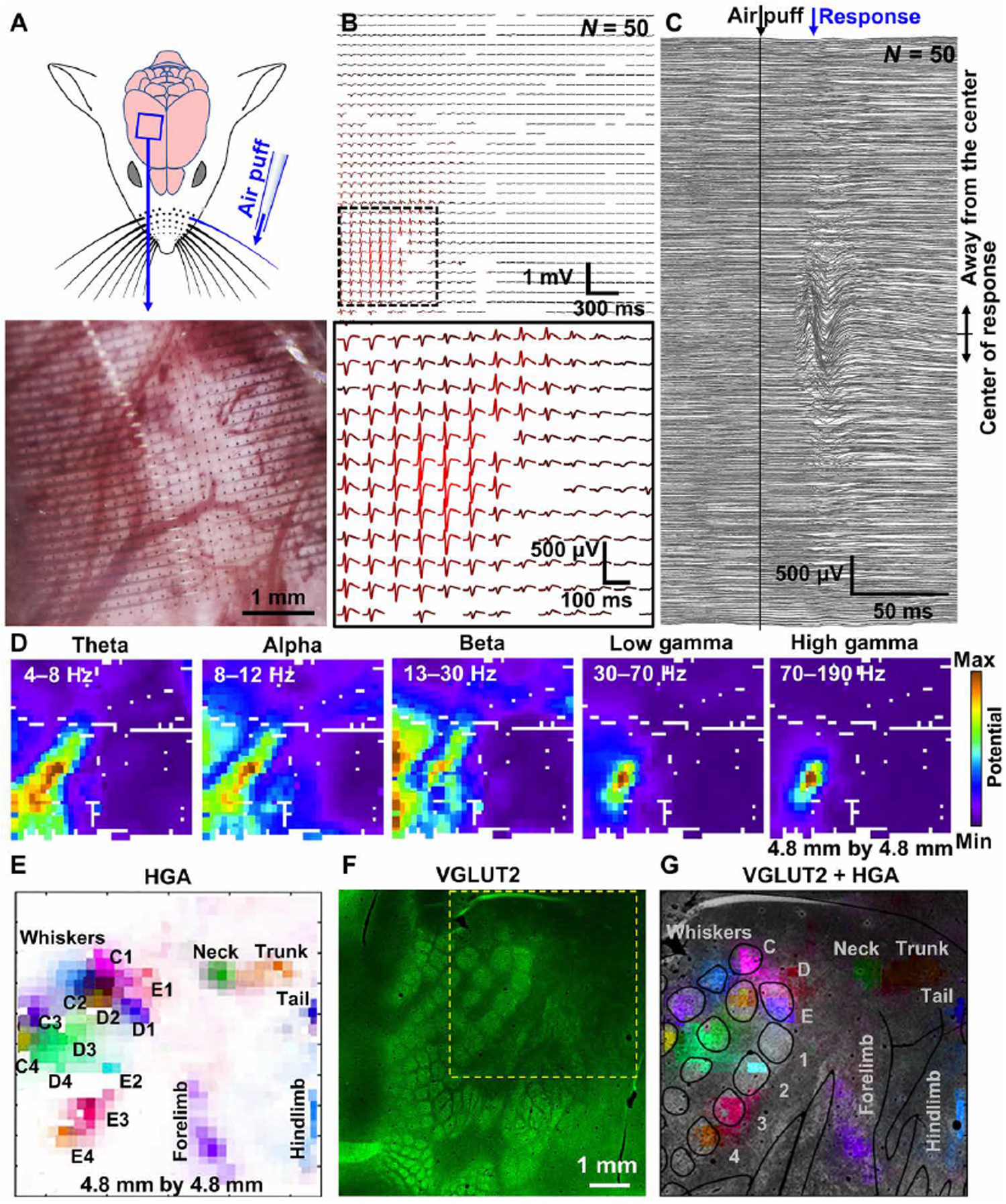
Fig. 2. PtNR multi-thousand channel ECoG grids record somatotopic functional cortical columns with sub-millimeter resolution.

(A) Schematic of the rat brain implanted with 1024 channels, 4.8 mm × 4.8 mm array, and the air-puff stimulation of individual whiskers. The lower image shows the magnified microscope image of the electrode on the rat barrel cortex. (B) E4 whisker stimulation-evoked ECoG recordings (N=50, raw). (C) Stimulation locked response of all channels. (D) Spatial mapping of neural wave amplitude filtered at different frequency windows. (E) Spatial mapping of high gamma activity recorded by the high-density PtNRGrid. Each label indicates the positions stimulated with air-puff. (F) VGLUT2 immunostaining of the rat barrel cortex. The electrode implantation location is marked with the yellow dotted box. (G) HGA superimposed on top of the histology image.
