Fig. 1.
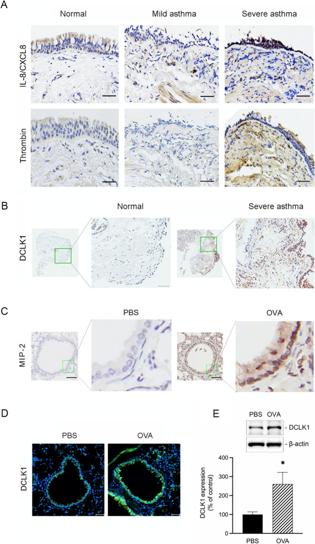
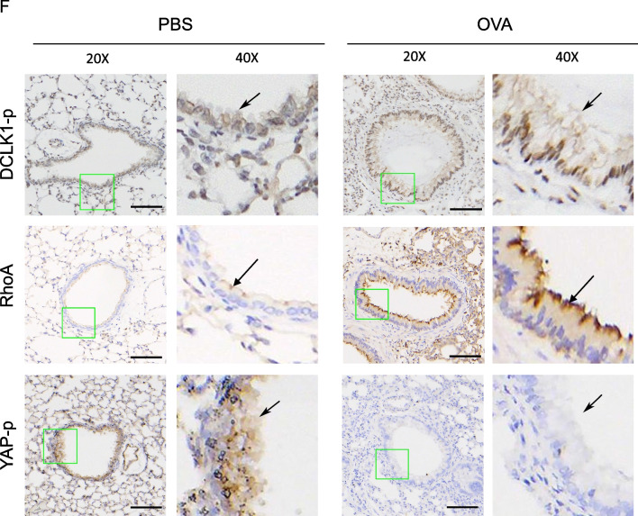
DCLK1 is overexpressed in the lungs of severe asthma patients and OVA-challenged mice. A Representative examples of IHC staining for IL-8/CXCL8 and thrombin in bronchial biopsies from severe asthma patients compared to normal subjects and from those with mild asthma (n = 4–6, original magnification = 20 × , bars = 100 μm). B IHC staining for DCLK1 in bronchial biopsies of severe asthma patients compared with normal subjects (n = 5, original magnification = ×20, bars = 50 μm). C Representative examples of IHC staining for MIP-2 in the lung tissues of PBS-treated and OVA-challenged mice (n = 6, original magnification = ×20, bars = 100 μm). D Lung tissues from PBS-treated and OVA-challenged mice were detected for the DCLK1 (green) and nuclei (blue). Merged fluorescence microphotographs were captured by immunofluorescence microscopy (n = 3, original magnification = ×20, bars = 50 μm). E Whole lung lysates from PBS-treated and OVA-challenged mice were immunoblotted with antibodies specific for DCLK1 and β-actin (n = 6, mean ± SEM, *p < 0.05 vs PBS group). F Representative examples of IHC staining for DCLK1 phosphorylation, RhoA, and YAP phosphorylation from the lung tissues of PBS-treated and OVA-challenged mice (n = 6, original magnification = ×20, bars = 100 μm)


