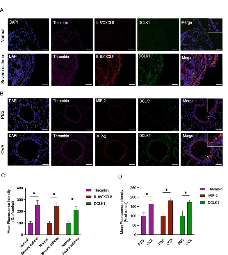
Fig. 2.
Triple immunofluorescence staining of thrombin, IL-8/CXCL8 (MIP-2), and DCLK1. A Representative examples of triple immunofluorescence staining for thrombin, IL-8/CXCL8, and DCLK1 in bronchial biopsies from severe asthma patients compared to normal subjects (n = 4–5, original magnification = ×20), and B OVA-challenged compared to PBS-treated mice (n = 5–7, original magnification = ×20). The merged image demonstrates colocalization of thrombin, IL-8/CXCL8, MIP-2, and DCLK1. The Image J software was used to measure the mean fluorescence intensity (MFI) of thrombin, IL-8/CXCL8 (MIP-2), and DCLK1 from C severe asthma patients compared to normal subjects (n = 4–5, mean ± SEM, *p < 0.05 vs normal) and D OVA-challenged compared to PBS-treated mice (n = 5–7, mean ± SEM, *p < 0.05 vs PBS)

