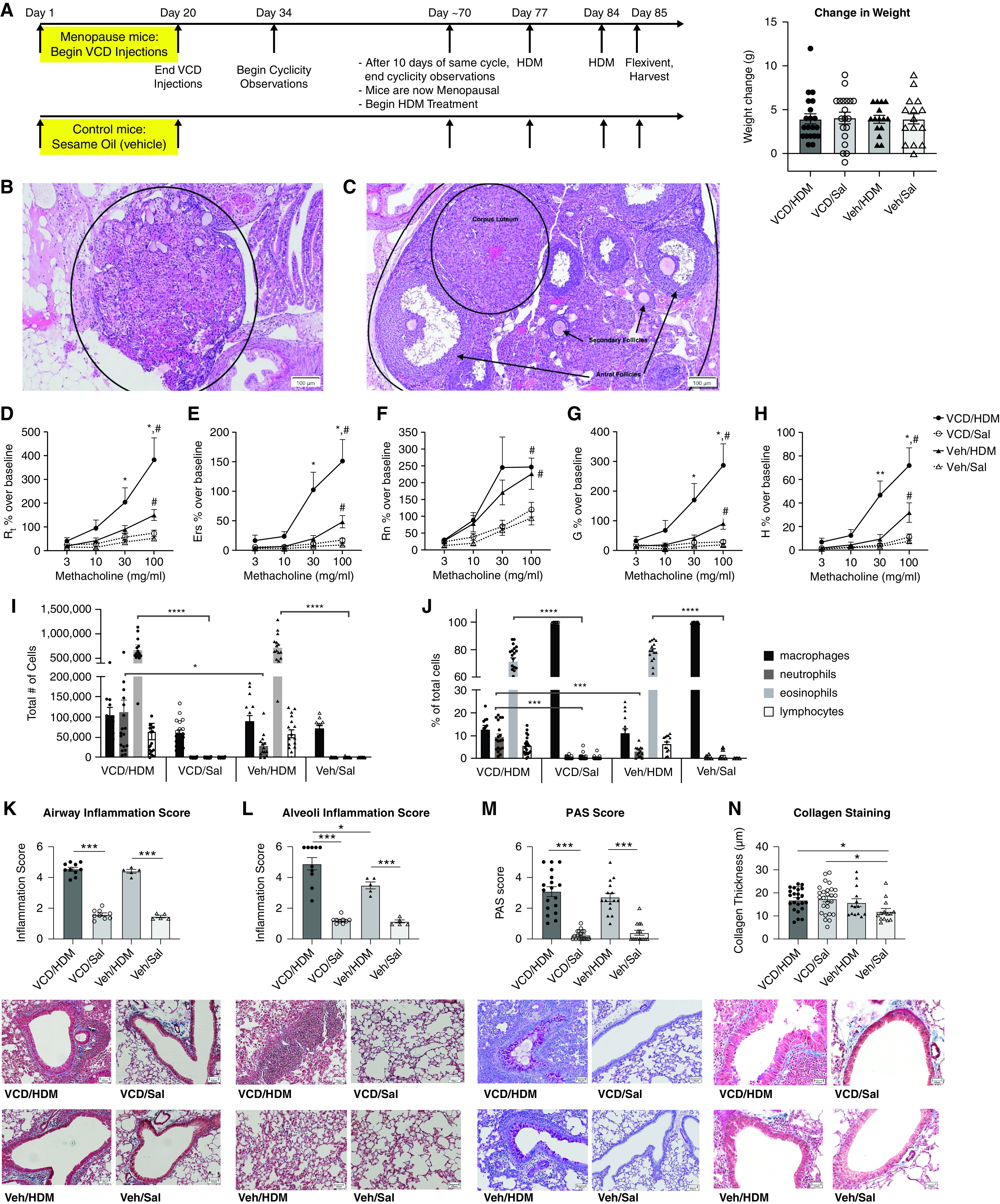
Figure 1.

Development of a novel menopause-associated asthma model in mice. (A) Experimental timeline for 4-Vinylcyclohexene diepoxide (VCD) injections and house dust mite (HDM) dosing, with the change in weight over the course of the experiment. (B) Hematoxylin and eosin (H&E) stained menopausal ovaries from VCD injected mice with follicle depletion as compared with (C) healthy ovaries from control mice that display follicles at various stages of development. Scale bar in B, 100 μm. Scale bar in C, 100 μm. (D–H) Lung function assessment in experimental mice. VCD/HDM (n = 20), VCD/Saline (n = 19), Vehicle/HDM (n = 15), and Vehicle/Saline (n = 15) female mice were assessed for pulmonary function 24 hours after the last HDM challenge and parameters are reported as percent over their respective baseline measurement; all HDM treated mice had significantly increased airway hyperresponsiveness (AHR) in all parameters of pulmonary functions assessed at the 100 mg/ml MCH dose compared with saline controls (#P < 0.05). D) Total airway resistance (Rrs): VCD/HDM mice compared with Veh/HDM mice at MCH doses: 30 mg/mL (*P = 0.0426) and 100 mg/mL (*P = 0.0334). E) Total airway elastance (Ers): VCD/HDM mice compared with Veh/HDM mice at MCH doses: 30 mg/mL (*P = 0.0101) and 100 mg/mL (*P = 0.0043). (F) Newtonian resistance (Rn): HDM-treated groups had significant increases in Rn compared with their respective saline controls (#P < 0.05). (G) Tissue damping (G): VCD/HDM mice compared with Veh/HDM mice at MCH doses: 30 mg/mL (*P = 0.0417) and 100 mg/mL (*P = 0.0281). (H) Tissue elastance (H): VCD/HDM mice had significantly higher H compared with Veh/HDM mice at MCH doses: 30 mg/mL (**P = 0.0026) and 100 mg/mL (**P = 0.0057). (I–J) The total number and percentage of inflammatory cells present in the BALF was determined by cytospin and differential counts after H&E stain. *P < 0.05, ***P < 0.001, and ****P < 0.0001 by one-way ANOVA. Lung sections from the right lobe were stained and assessed for inflammation, collagen, and mucin production at 10X microscopy. Masson’s Trichrome stained sections were assessed for (K) inflammation surrounding the large airways on a scale of 1 (no inflammation) to 5 (full inflammation) and (L) alveolar inflammation on a scale of 1 (no inflammation) to 6 (full inflammation). (M) PAS-stained sections were analyzed for mucin production using a scale of 0 (no mucin) to 5 (full mucin production). (N) Masson’s Trichrome stained sections were assessed for collagen (blue) thickness surrounding the large airways using MetaMorph software. *P < 0.05 and ***P < 0.001 by one-way ANOVA for multiple comparisons. Veh = vehicle.
