
Figure 1. Examples of New or Enlarging Lesions Occurring Between Attacks in Myelin Oligodendrocyte Glycoprotein Antibody-Associated Disease.
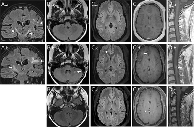
(A) The reference coronal MRI (T2-FLAIR) image reveals bilateral internal capsule and a left hemispheric T2-hyperintense lesion (A1, arrowheads) that on follow-up showed enlargement of the right internal capsule and left subcortical white matter T2-hyperintense lesions (A.b, arrows) in the absence of a new attack. (B) The reference axial MRI T2-FLAIR image (B.a) reveals normal brainstem and cerebellum signal while the follow-up image shows a new T2-hyperintensity in the left middle cerebellar peduncle (B.b, arrow) in the absence of a new clinical attack. The T2-lesion resolved completely and was no longer visible on a subsequent MRI FLAIR image (B.c) highly consistent with the expected evolution of a MOGAD lesion. (C) The reference axial MRI T2-FLAIR image (C.a) and axial T1 postgadolinium image (C.b) of the supratentorial region reveals no abnormalities but on follow-up show a new T2-hyperintensity had developed in the right frontal region (C.c, arrow) that enhanced after gadolinium (C.d, arrow) in the absence of a new attack. The lesion had resolved completely and was no longer visible on FLAIR (C.e) or T1 postgadolinium (C.f) images on a subsequent MRI highly consistent with the expected evolution of a MOGAD lesion. (D) The reference sagittal MRI cervical spine T1-weighted images postgadolinium revealed some subtle gadolinium enhancement (D.a, arrowhead) that increased in size in the follow-up (D.b, arrow) in the absence of a new attack despite no change in the T2-hyperintense cord lesion (not shown). The enhancement had resolved completely and was no longer visible on subsequent T1 postgadolinium image (D.c) highly consistent with the expected evolution of a MOGAD lesion. Abbreviations: FLAIR = fluid attenuated inversion recovery; MOGAD = myelin oligodendrocyte glycoprotein antibody-associated disease.