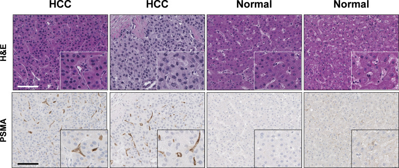
Fig 2. Representative immunohistochemical staining for prostate-specific membrane antigen (PSMA) in formalin-fixed paraffin-embedded liver tissues.
PSMA expression was assessed using anti-PSMA antigen antibody (Cell Signaling, Danvers, MA; D718E clone) and avidin-biotin peroxidase immunohistochemistry. Hematoxylin and eosin staining of formalin-fixed paraffin-embedded human liver samples diagnosed as normal and hepatocellular carcinoma (HCC) are shown at the top of the image. High magnification images are shown in the inset. Scale bar shown is 100 μm. These images were created by co-author Joon-Yong Chung at the National Cancer Institute, with permission privileges for their use.

