
FIGURE 2.
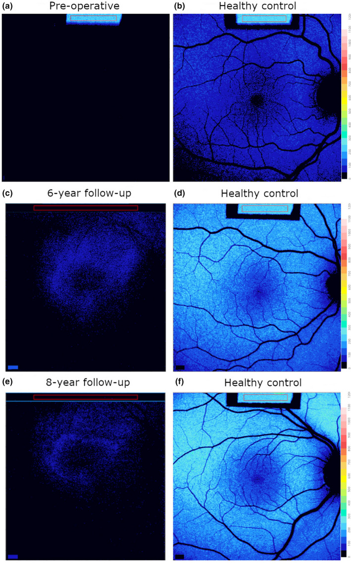
Quantitative fundus autofluorescence (qAF) color‐coded images in the patient prior to treatment and at 6‐ and 8‐year follow‐up. Right eyes. (a,b) qAF prior to intervention; near absence of autofluorescence (a) relative to a healthy control (b). (c,d) qAF image revealed parafoveal autofluorescence at 6‐year follow‐up (c) compared with healthy age‐matched control (d). (e,f) 8‐year (e) follow‐up compared with control (f).