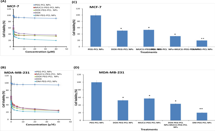
Fig. 4.
Impact of NPs on DOX and MUC1 inhibitor intracellular location and cell viability. MCF-7 (A) and MDA-MB-231 (B) cells were treated at 1:1 ratios with NPs entrapped with DOX and MUC1 inhibitor.MTT tests were used to examine the cells after 48 h. The % viability (mean ± SD of three separate trials) is provided. MCF-7 (A) and MDA-MB-231 (B) cells were given 48 h of treatment with PEG-PCL NPs, DOX-PEG-PCL NPs, MUC1i-PEG-PCL NPs, and DM-PEG-PCL NPs. Cell viability was determined using MTT assay. The % viability (mean ± SD of three separate trials) is provided. MCF-7 (C) and MDA-MB-231 (D) cells were treated for 72 h with a 0.01 µM concentration each. MTT tests were used to evaluate cell viability. The results are given as % of viable cells. (mean ± SD of three replicates)

