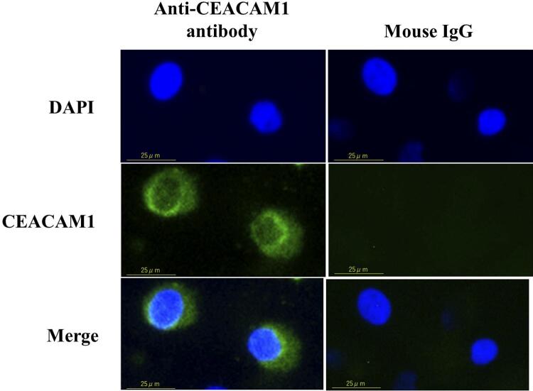
Figure 1. Expression of CEACAM1 in oral keratinocytes and fibroblasts.
Localization of CEACAM1 expression in RT7 cells. Cells were stained with anti-CEACAM1 or mouse IgG as a negative control along with Alexa Fluor® 488 conjugated mouse IgG, and nuclei were counter-stained with DAPI (blue). Green staining indicates CEACAM1. Each experiment was performed at least three times, with representative results shown.

