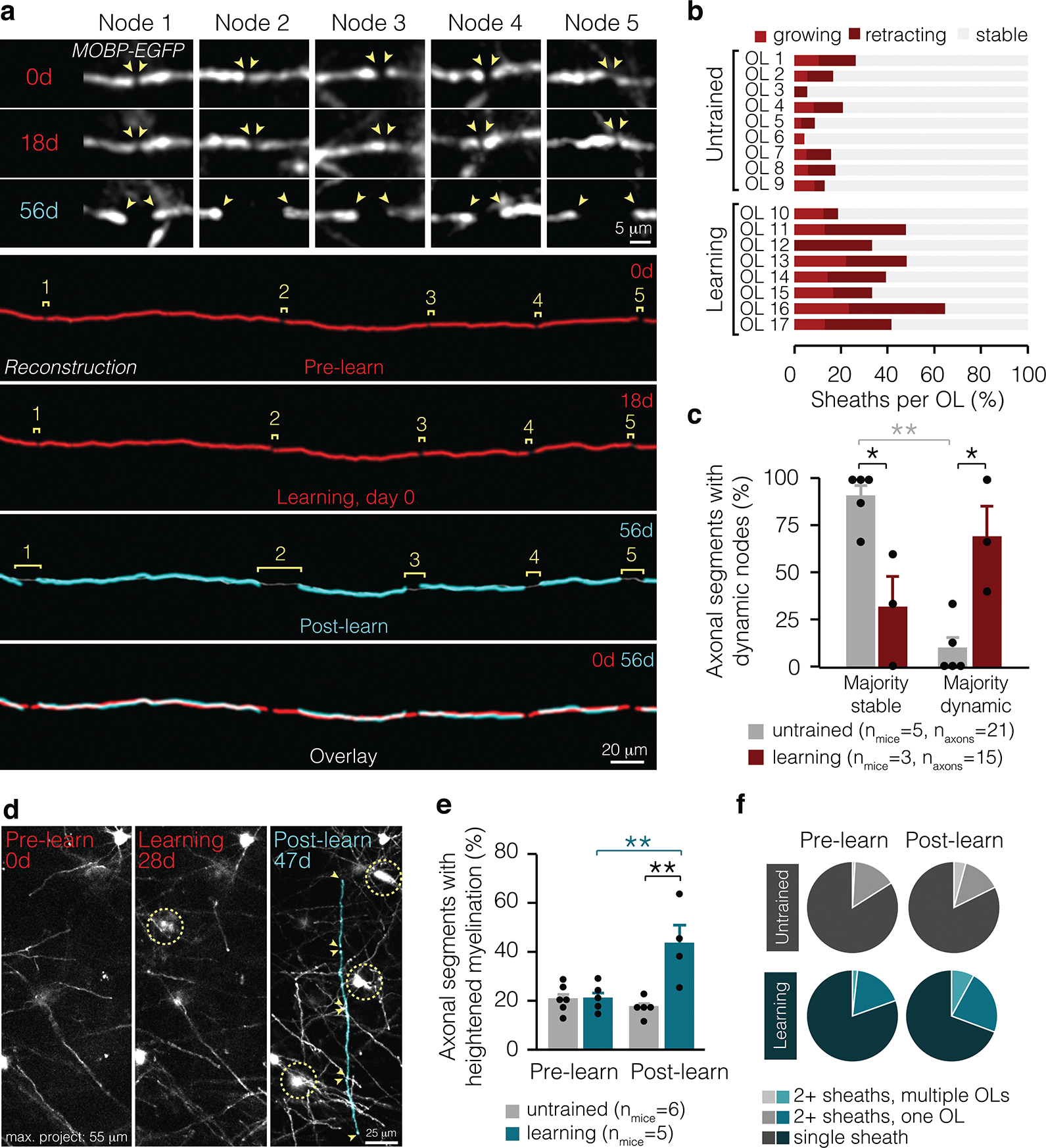
Figure 3 |. Learning-induced changes to myelin occur at specific locations along axons.

a, Multiple nodes along the same axonal segment lengthen in response to learning (in vivo data shown on top, reconstructions shown on bottom). b, Individual oligodendrocytes in learning and untrained mice exhibit multiple sheath behaviors. c, Learning modulates the proportion of axonal segments with majority dynamic nodes (F3,12 = 14.17, p = 0.0003). In learning mice, significantly more axonal segments with dynamic myelin possess majority dynamic nodes than in untrained mice (p=0.01; Tukey’s HSD). d, Axons can be myelinated by multiple new sheaths. Sheaths of interest are pseudo-colored. e, Learning modulates the proportion of axonal segments receiving multiple new myelin sheaths (F1,9.46 = 14.17, p = 0.0027). In the two weeks following learning, axons are more often myelinated by multiple new sheaths than before learning (p = 0.0055; Tukey’s HSD). The percent of axonal segments receiving increased myelin sheaths is heightened in learning compared to untrained mice (p = 0.006; Tukey’s HSD). f, Proportion of axonal segments receiving new myelin from multiple oligodendrocytes, multiple sheaths from a single oligodendrocyte, or only one new sheath in untrained mice (gray) and before and after learning (blue). *p<0.05, **p < 0.01, ***p < 0.0001, NS, not significant; bars and error bars represent mean ± s.e.m. For detailed statistics, see Supplementary Table 3, Figure 3.
