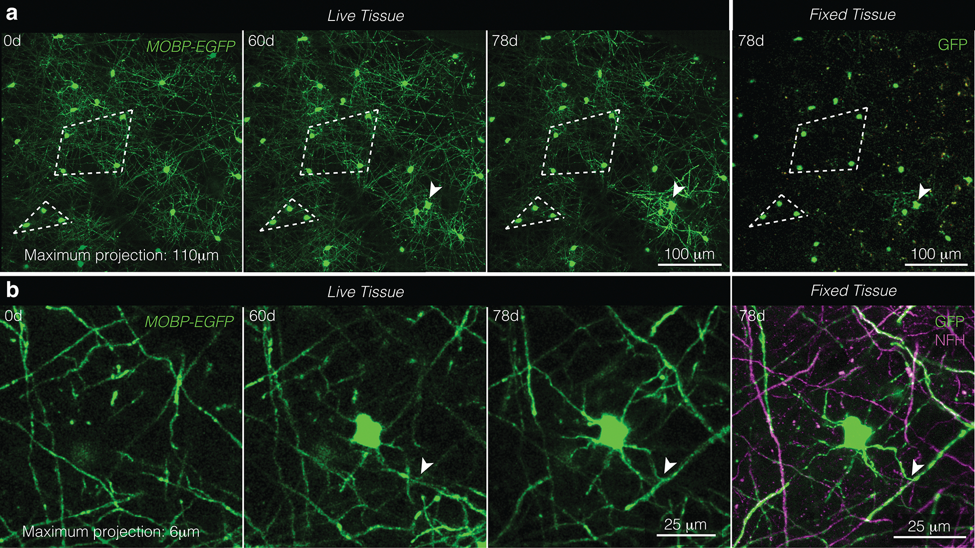
Extended Data Fig. 1. Near-infrared branding identifies the same oligodendrocytes and myelin sheaths in longitudinally, in vivo imaged areas and post-hoc stained tissue.

a, The same field of view imaged in vivo (“Live Tissue”, left) and fixed, sectioned, and stained tissue (“Fixed Tissue”, right). Patterns of cell bodies (examples outlined in white dotted lines) were maintained across live and processed tissue. Note the new oligodendrocyte generated at 60d and delineated with a white arrowhead. b, A newly generated oligodendrocyte in vivo (“Live Tissue”, left) and fixed, sectioned tissue (“Fixed Tissue”, right) stained for oligodendrocytes and myelin (GFP, green) and axons (NFH, magenta). Note the same T-junction across live and fixed samples is marked with the white arrowhead.
