Figure 1.
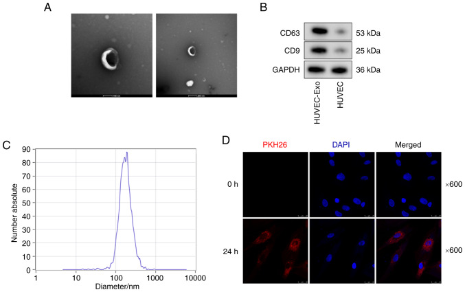
Exosomes were identified and taken up by VSMCs. (A) Transmission electron microscopy was used to assess the morphology of exosomes. Scale bar, 100 nm (left) or 200 nm (right). (B) Exosome-specific markers were detected by western blot analysis. (C) Nanoparticle tracking analysis of exosomes derived from VSMCs showed that the size of most HUVEC-Exos was 80-170 nm. (D) VSMCs were incubated with PKH26 fluorescently labeled exosomes for 0 and 24 h. Confocal microscopy analyses was used to identify the uptake of exosomes by VSMCs (PKH26 in red; DAPI in blue). Magnification, ×600. HUVEC-Exos, exosomes from HUVECs; VSMC, vascular smooth muscle cell.

