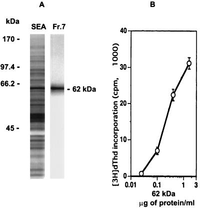
FIG. 3.
Identification of a 62-kDa antigen recognized by T-cell hybridoma 4E6. (A) Eluted SDS-PAGE gel fraction 7 (Fr.7) (Fig. 1) from 20 μg of SEA, examined for purity on a 10% silver-stained SDS-polyacrylamide gel, shown next to total SEA and molecular weight marker standards. (B) Dose response of T-cell hybridoma 4E6 to 62-kDa antigen as measured by HT-2 indicator cell proliferation. Data are expressed as mean ± 1 SD. Background radioactivity from hybridoma cultures in the absence of antigen is subtracted.

