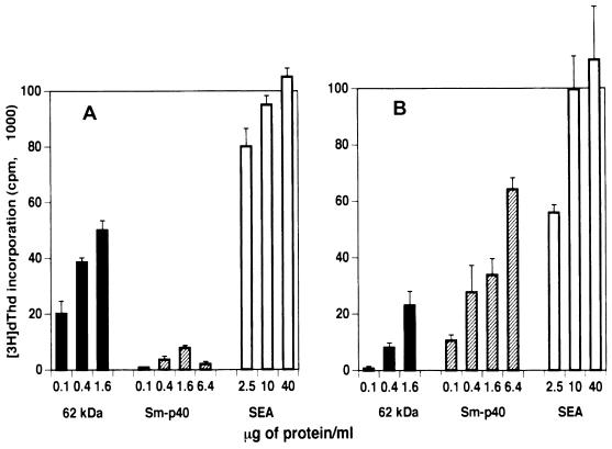
FIG. 6.
Proliferative responses of CD4+ Th cells from BL/6 (A) and CBA (B) mice to the 62-kDa antigen. CD4+ Th cells were isolated from mesenteric lymph nodes of 8.5-week-infected mice. Culture conditions were as described in Materials and Methods. [3H]dThd incorporation was assessed by liquid scintillation spectroscopy. Data are expressed as mean ± 1 SD. Also shown for comparison are responses to Sm-p40 and SEA. Background radioactivity from cultures in the absence of antigen is subtracted. The same pattern of stimulation was observed when cells from 7.5-week-infected mice were used (not shown).

