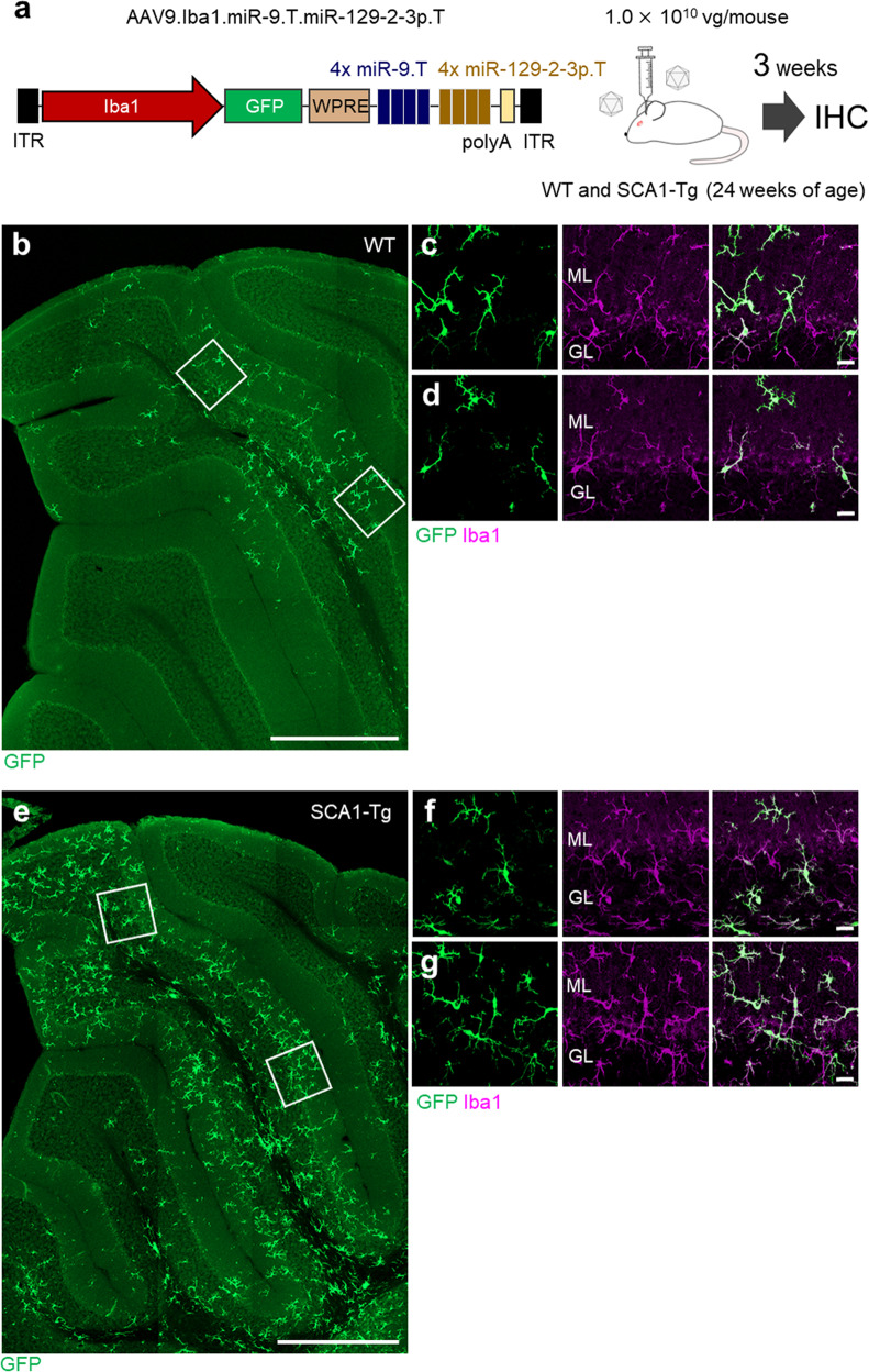
Fig. 5. Robust transduction of microglia in symptomatic SCA1-Tg mouse cerebellum.
a AAV construct (AAV9.Iba1.miR-9.T.miR-129-2-3p.T) and schematic of the virus injection. AAV (dose: 1.0 × 10¹⁰ vg/mouse) was injected into the wild-type and SCA1-Tg mouse cerebellum at 24 weeks of age. Three weeks after the viral injection, cerebellar sections were analyzed by immunohistochemistry. b–d GFP immunofluorescent image of the wild-type (WT) mouse cerebellum. e–g GFP immunofluorescent image of a sagittal section of the SCA1-Tg (B05) mouse cerebellum. Square regions in b and e were enlarged to c, d and f, g, respectively. Scale bars; 500 μm (b, e) and 20 μm (c, d, f, g). GL granule cell layer, ML molecular layer.

