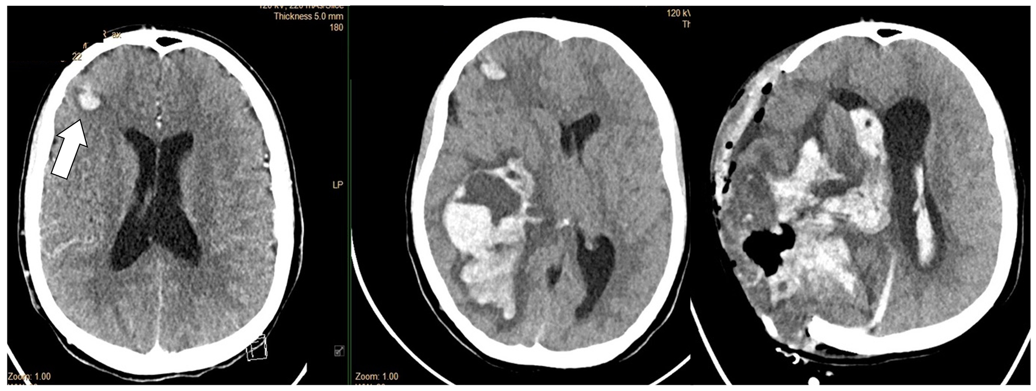
Figure 6.

Adolescent with DiGeorge Syndrome and repaired truncus arteriosus with St. Jude’s valve in the mitral position and infective endocarditis. The head CT demonstrated multiple septic emboli with hemorrhagic transformation (small right frontal lesion demonstrated on this slice) (left panel). Head CT 48 hours later with massive hemorrhage before (middle panel) and after (right panel) craniectomy. CTA was not performed, but the large hemorrhage was presumed to be due to a ruptured infectious intracranial aneurysm.
