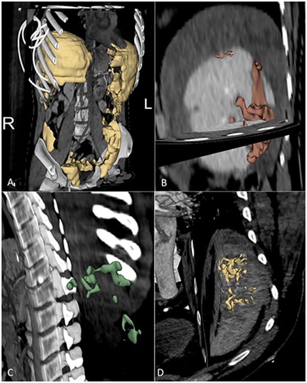
Figure 1.
Voxelwise multiclass labels of CT features associated with splenectomy and angioembolization in two patients with splenic injuries. A-C: 22-year-old male with BSI after motor vehicle collision. A volume rendered PV phase image shows segmented hemoperitoneum (yellow, Part A) throughout the abdomen and pelvis, with a total volume of 2000 mL. Sagittal oblique PV phase images in the same patient show a splenic laceration (red, Part B), measuring 71 mL, and foci of active hemorrhage (green, Part C), with volume totaling 9 mL. D: 55-year-old male status post fall. An oblique maximum intensity projection image from admission contrast enhanced arterial phase CT shows extensive pseudoaneurysms (yellow, Part D), with a total volume of 8 mL.

