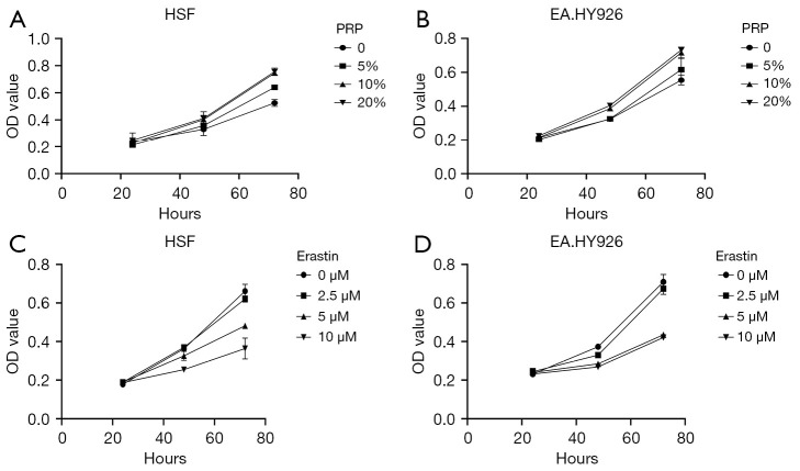
Figure 1.
PRP and inhibition of ferroptosis in HSF and EA.HY926 cells exposed to HG conditions. (A,B) The CCK-8 detection results of PRP on HSF and EA.HY926 cells. It can be seen that within the same culture time range, with the increase of PRP, the cell survival rate showed an increasing trend. Under the intervention of the same drug concentration, the cell survival rate increased with the prolongation of the treatment time of PRP. (C,D) The effect of Erastin on the survival rate of HSF and EA.HY926 cells. It can be seen that the survival rate of cell decreases with the extension of the duration of Erastin when the drug concentration is fixed. With the increase of Erastin concentration, the survival rate of fibroblast HSF decreased. OD, optical density; PRP, platelet-rich plasma; HSF, human skin fibroblasts; HG, high glucose.

