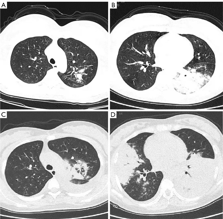
Figure 4.
Representative CT images of SMPP in pregnant women. (A,B) Multilobar infiltration and pleural effusion were shown on different levels. (C,D) One week later, the same level of imaging showed deteriorated multilobar infiltration, atelectasis, and increased pleural effusion. CT, computed tomography; SMPP, severe Mycoplasma pneumoniae pneumonia.

