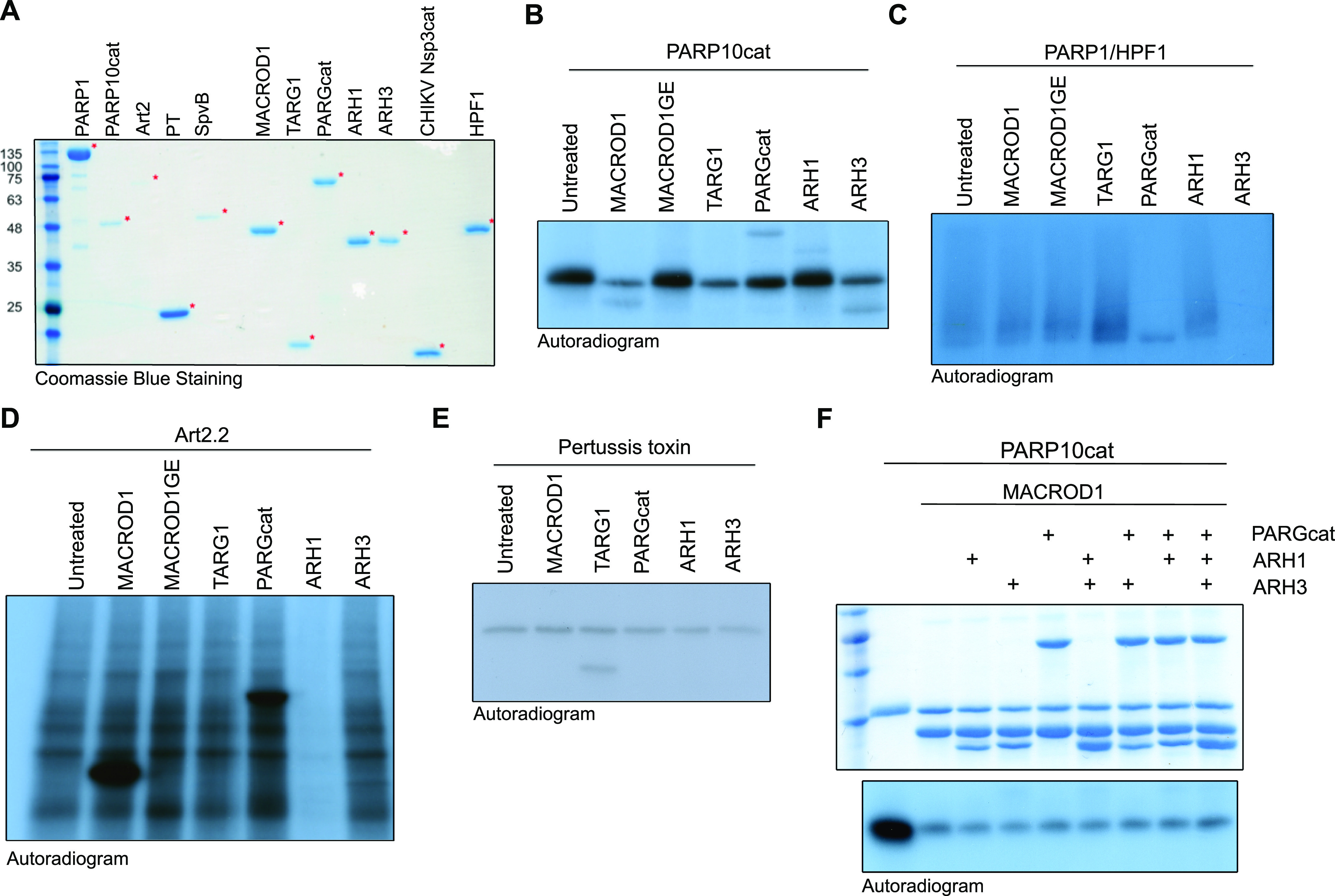
Figure 1. Enzymatic generation of specific ADP-ribosylated substrates.
(A) Coomassie blue staining of the purified transferases, hydrolases, and co-factors used in this study. (B, C, D, E) Indicated transferases were incubated with 32P-NAD+ and incubated at 37°C for 30 min. A cytoplasmic extract was provided to supply the mAART2.2 and pertussis toxin substrates. After the transferase reaction, OUL35 was added to inhibit PARP10 or olaparib was added to inhibit PARP1/HPF1, followed by a 30-min hydrolase reaction. Samples were run on SDS–PAGE, and incorporated radioactivity was visualised. The indicated MACROD1 mutant is an inactivating glycine 270 to glutamate mutation. (F) PARP10 was automodified using 32P-NAD+, the reaction was stopped with OUL35, and indicated hydrolases were added. The Coomassie staining is displayed to show the relative amounts of hydrolases added. The samples were analysed as in (B).
Source data are available for this figure.

