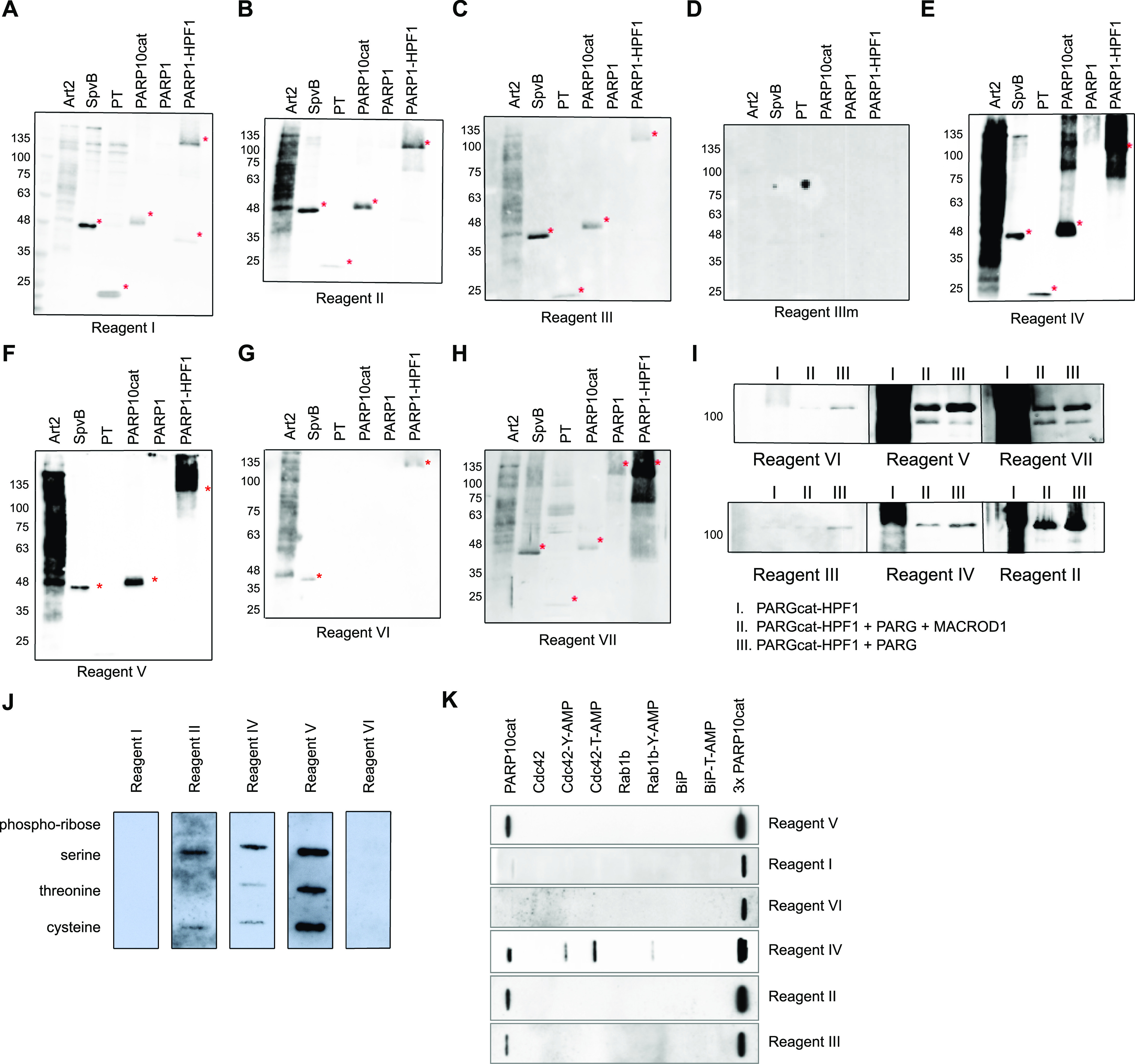
Figure 2. Specificity of the reagents towards in vitro modified substrates.
(A, B, C, D, E, F, G, H) ADP-ribosylation reactions from Fig S1 were loaded on multiple gels and blotted. Membranes were blocked with 5% milk in TBST and incubated with primary antibodies overnight. Asterisks indicate the transferases. (I) GFP-PARP1/His-HPF1 was incubated with NAD+ (I), followed by PARGcat and MACROD1 (II) or PARGcat (III) treatment. After blotting, the modification was detected using the same reagents. (J) 2 µM chemically synthesised peptide modified with either phospho-ribose or ADP-ribose on serine, threonine, or cysteine was slot-blotted and analysed using the indicated reagents. (K) 150 ng of AMPylated proteins was slot-blotted and analysed using the indicated reagents.
Source data are available for this figure.

