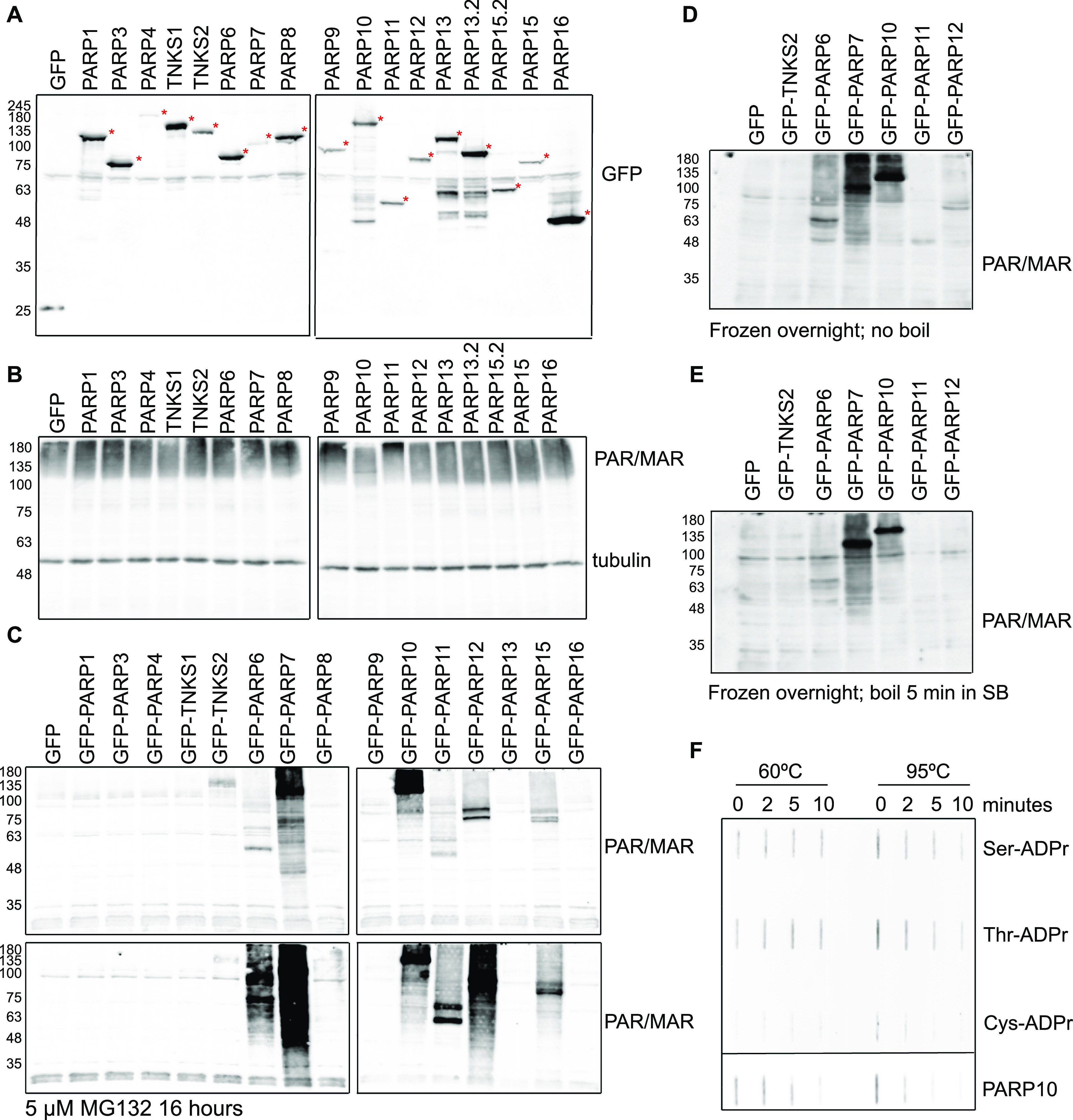
Figure 3. Optimised lysis conditions are required to prevent ADP-ribosylation from occurring during lysis or degradation during sample preparation.
(A) HEK293T cells were transfected with indicated GFP-PARPs, lysed in RIPA buffer, and analysed using Western blotting with a GFP antibody. (B) Same lysates as in (A), but analysed with ADPr antibody Reagent V. Subequently the blot was detected using a tubulin antibody. (C) HEK293T cells were transfected with indicated GFP-PARPs and lysed in RIPA buffer supplemented with olaparib or in addition treated with proteasomal inhibitor MG132 before lysis. Western blots were analysed with ADPr antibody Reagent V. (D, E) Untreated lysates from (C) were frozen at −20°C and heated at (D) 60°C or (E) 95°C before loading the SDS–PAGE. Resulting Western blots were analysed with ADPr antibody Reagent V. (F) ADP-ribosylated peptides and automodified PARP10 catalytic domain were slot-blotted, untreated, or heated at 60°C and 95°C for 2, 5, or 10 min. The blot was analysed using an ADPr antibody Reagent V. The PARP10 blot was exposed shorter because of the stronger signal; different exposures are provided as source data.
Source data are available for this figure.

