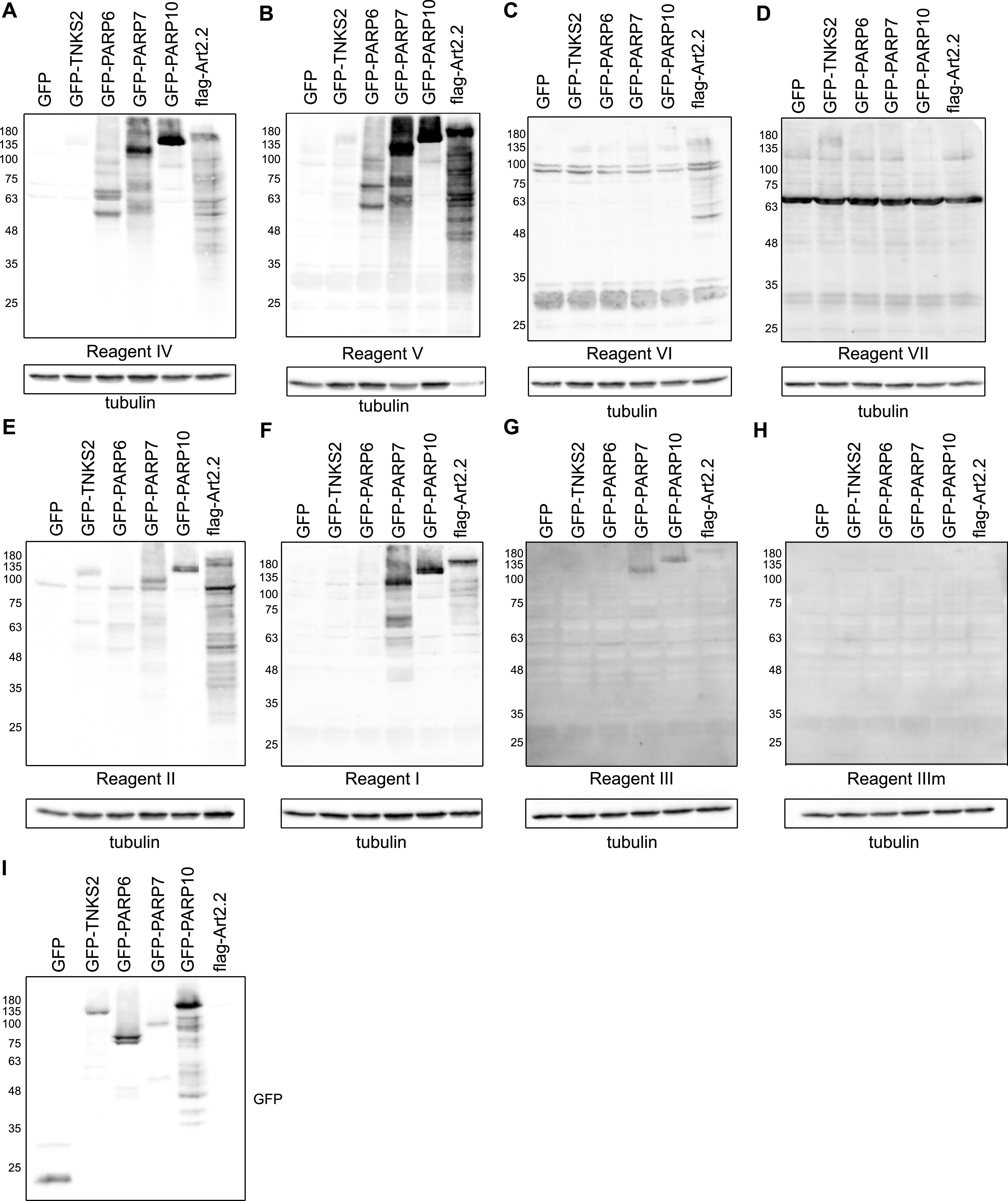
Figure 4. Anti-ADP-ribose detection reagents have different specificities for substrates modified in cells.
(A, B, C, D, E, F, G, H) HEK293T cells were transfected with the indicated GFP-tagged PARP constructs, GFP as control or flag-tagged murine ART2.2. 24 h after transfection, cells were lysed in RIPA buffer supplemented with olaparib and analysed using SDS–PAGE. The Western blot was detected with the indicated reagents. (I) Western blot showing the transfection levels of the GFP-transferases.
Source data are available for this figure.

