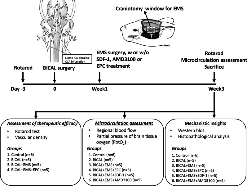
Fig. 1.
Schematic representation of the experimental procedure. BICAL surgery was performed by permanent ligation of the ICA distal to the bifurcation of the CCA. One week following BICAL surgery, the craniotomy window for EMS (white dotted box) was created after downward reflection of the temporalis muscle (marked as T). Treatment with EPCs, SDF-1 or AMD3100 was administered during the EMS procedure. The rotarod test was performed 3 days before and 3 weeks after BICAL. Vascular density and microcirculation were evaluated 3 weeks after BICAL. Animals were killed 3 weeks following BICAL surgery, and brain tissues were harvested for Western blot analysis and histopathological analysis

