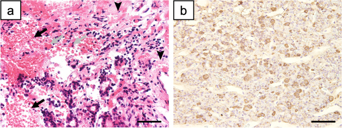
Fig. 2.
Histopathological examination of the pituitary mass removed on day 52. a Hematoxylin & eosin-stained image. The arrowheads indicate necrotic findings in the glandular pituitary region. The black arrows indicate hemorrhage, with accumulation of erythrocytes outside the blood vessels. The grey arrows indicate macrophages, with phagocytosis of red blood cells. b Anti-ACTH-immunostained image. The reddish-brown area is ACTH-positive, indicating that the resected pituitary tissue is an ACTH-producing adenoma. Scale bars: 20 μm. ACTH: adrenocorticotropic hormone

