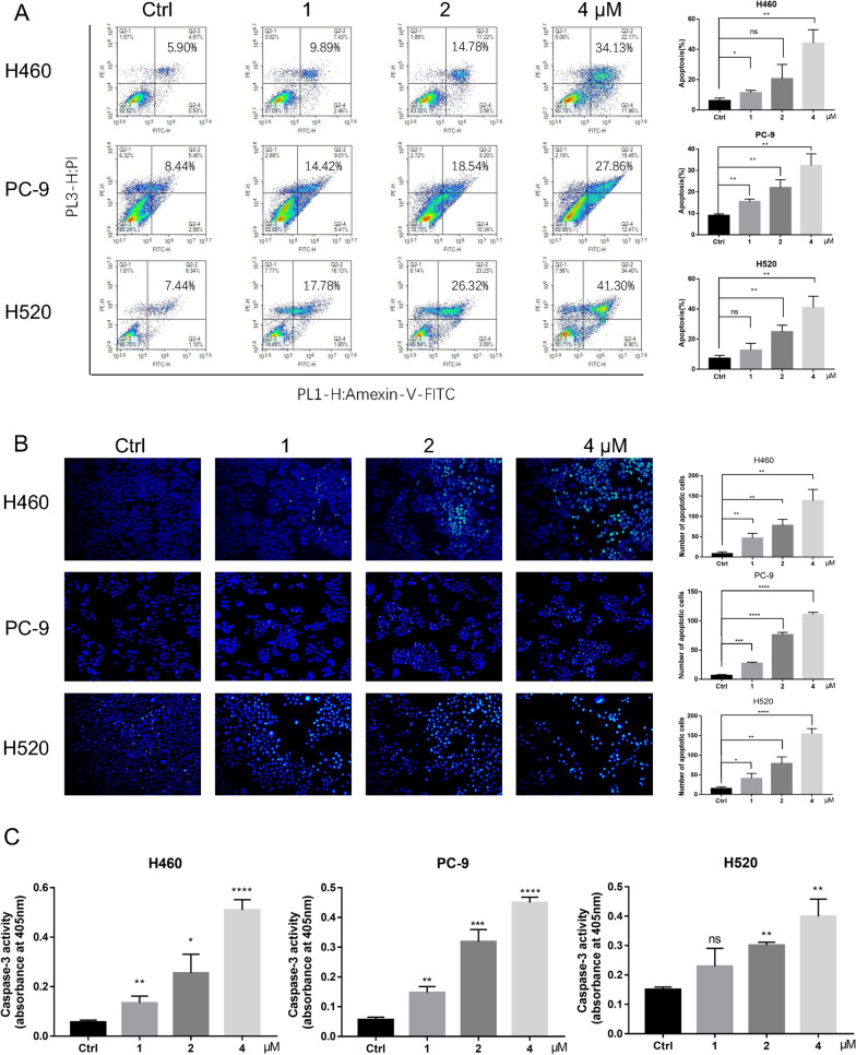
Fig. 2.
Celastrol induced apoptosis of human NSCLC Cells. A After treatment with celastrol at the indicated concentration for 24 h before staining with Annexin V and propidium iodide (PI), the distribution of apoptotic cells was determined using flow cytometry. B The apoptotic characteristics in NSCLC cell nucleus presented by Hoechst Staining assay. After treatment with celastrol for 12 h before staining with Hoechst staining, the nucleus was observed and photographed (100×). Representative results of triple experiments. C Caspase 3 activity was evaluated using the Caspase Assay Kit, and absorbance was recorded at 405 nm. (*P < 0.05, **P < 0.01, ***P < 0.001, ****P < 0.0001)

