Fig. 5.
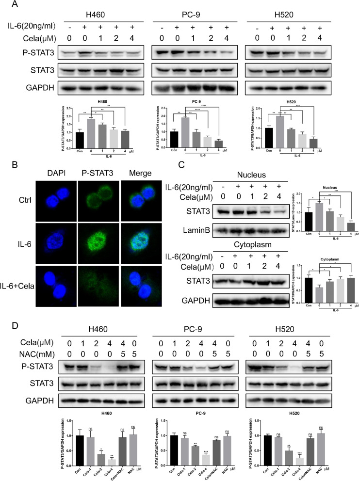
Celastrol inhibited the IL-6/STAT3 signaling pathway in NSCLC cells. A H460, PC-9, and H520 cells were exposed to IL-6 for 30 min after treatment with celastrol for 3 h, and the expression level of P-STAT3 was detected by western blotting. B Representative confocal microscopic images indicating the localization of p-STAT3 (green) and DAPI in H460 cells. C The expression levels of STAT3 in nuclear and cytosolic fractions were determined by using the western blot analysis. Images shown are representative of three separate experiments. D H460, PC-9, and H520 cells were treated with celastrol or in combination with NAC. The expression of p-STAT3 and STAT3 was detected by western blotting. Outcomes are representative of three independent experiments (*P < 0.05, **P < 0.01, ***P < 0.001, ****P < 0.0001)

