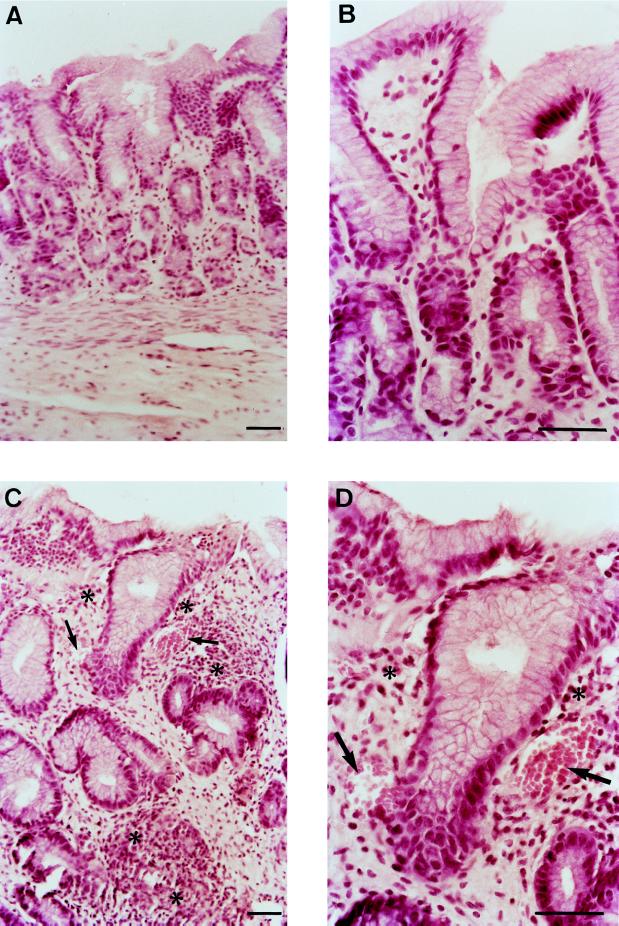
FIG. 2.
Hematoxylin-and-eosin-stained sections of human gastric mucosa from uninfected and infected xenografts at 12 weeks after inoculation with H. pylori LB1. (A and B) Normal gastric antral mucosa. (C and D) Gastric infected mucosa, showing dilated capillaries (arrows) and mild infiltration of mononuclear cells and rare polymorphonuclear leukocytes (∗). Bars = 50 μm.

