Figure 3.

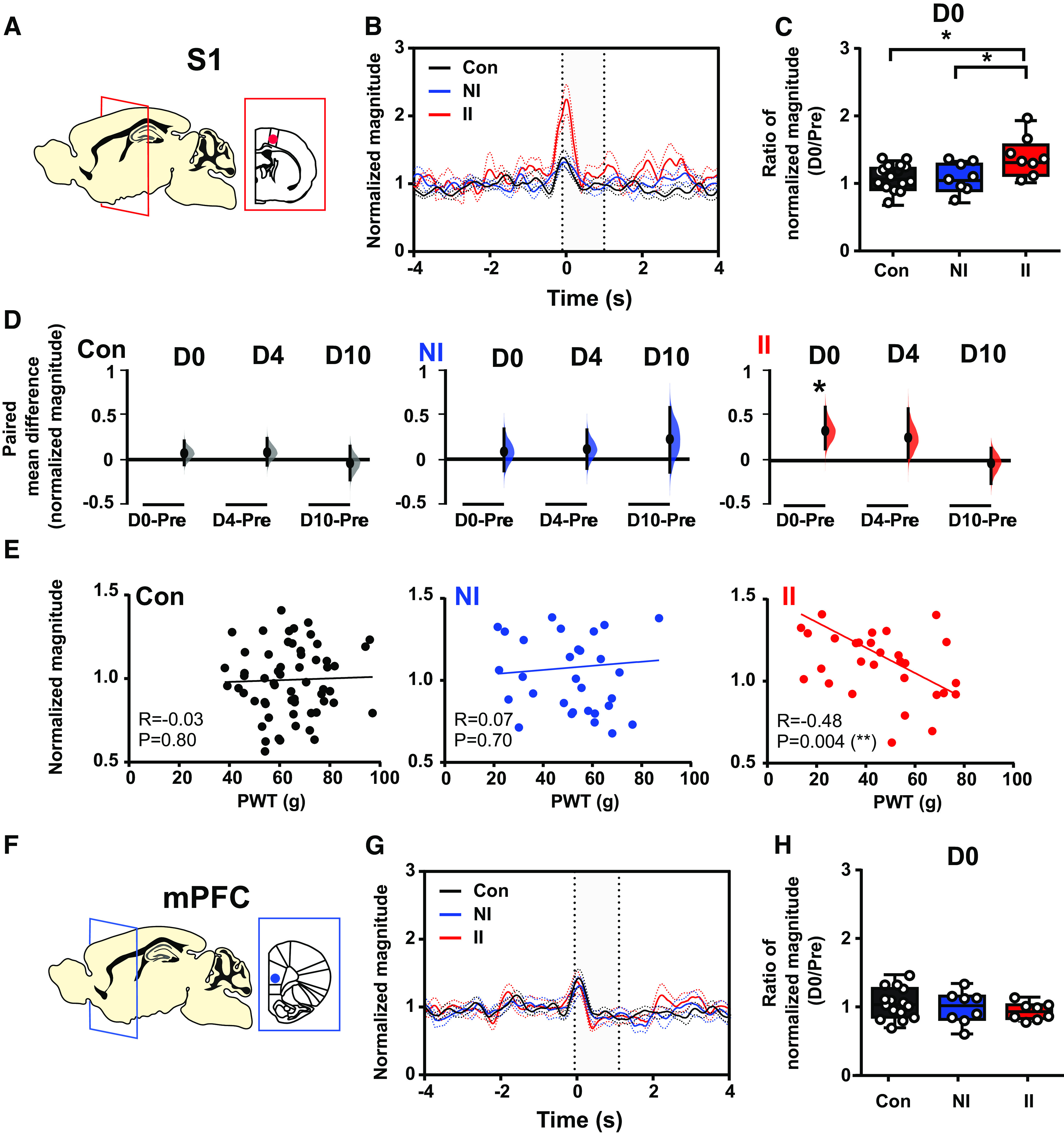
Stimulus-evoked δ energy in SI increases after adult incision injury only in animals who experienced ELP. Electrophysiological responses in the (A) somatosensory cortex (S1) and (F) mPFC to mechanical (eVF) stimulation of the hindpaw following adult injury in ELP rats (II, red) and non-ELP rats (NI, blue) and controls (Con, black). Peristimulus normalized δ frequency (2–4 Hz) oscillations (mean ± SEM) in S1 (B) and mPFC (G) on the day of adult incision injury (D0). Comparison of the injury-induced changes in stimulus-evoked δ energy in S1 (C) and mPFC (H), expressed as a ratio of normalized magnitude (D0/Pre), between groups. D, The enhancement of injury-induced changes in sensory evoked S1 δ energy returned to preinjury level by 10 d (D10) following injury. The paired mean difference for comparisons is shown as Cumming estimation. Each paired mean difference is plotted as a bootstrap sampling distribution; 95% confidence intervals are indicated by the ends of the vertical error bars. Statistical analysis was performed using a permutation t test (randomization: 5000). E, Correlations between PWT and stimulus-evoked S1 δ activity (normalized magnitude). The scatter plots represent the correlations between PWT and normalized energy (Pre to D10) with continuous lines showing the linear regression. Pearson correlation coefficient (R) with significance (p value) is presented in the figures. Nonincised adult controls (Con, n = 15), incision in adults without neonatal incision (NI, n = 8), and incision in adults with neonatal incision (II, n = 8).
