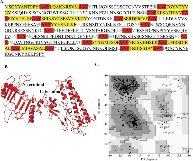
Figure 2.
Modeled structure of multi-epitope protein against LSDV. (A) Protein sequence of multi-epitope protein. Sequence in underline: B-cell epitope; yellow color: CTL epitope; green color: extra sequences for the stability of multi-epitope protein; Highlight red color: AAY linker, (B) Three-dimensional model of multi-epitope protein obtained by modeling and refinement by RaptorX. The gray color represents AAY linker, (C) Ramachandran plot analysis of the multi-epitope protein showing favored (80.3%), allowed (16.3%), generously allowed (1.9%), and disallowed regions (1.5%).

