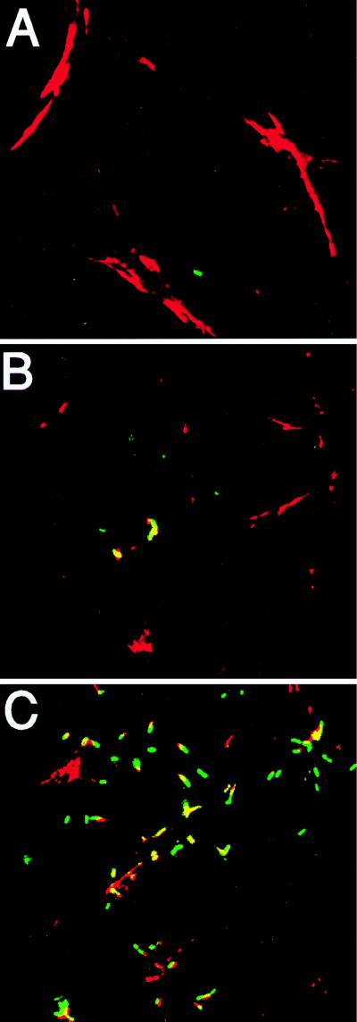
FIG. 4.
Intracellular fluorescence and F-actin staining of L. monocytogenes-infected PtK2 cells. Monolayers of PtK2 cells were grown on coverslips and infected with the NF-L357 actA-gfp-plcB fusion strain. Coverslips were removed at 3 h postinfection (A), 3 h postinfection (B) (an independent field adjacent to that shown in panel A), and 6 h postinfection (C) and treated with rhodamine phalloidin to identify F-actin. The yellow areas indicate overlapping fluorescence of GFP (green fluorescence) and F-actin (red fluorescence).

