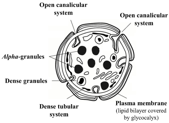
Figure 2.
Schematic image of platelets. Resting platelets have asymmetrical distribution of phospholipids: the anionic phospholipids phosphatidylserine and phosphatidylethanolamine, responsible for binding many of the blood coagulation proteins, are sequestered in the inner leaflet of the membrane lipid bilayer facing the cytosol, whereas the electrically neutral phosphatidylcholine and sphingomyelin are exposed on the outer membrane leaflet. This arrangement prevents the membranes of resting platelets from supporting coagulation. When platelets become activated, the function of the phospholipid transporters is altered, leading to transfer of anionic phospholipids on the outer membrane leaflet. Loss of this asymmetry provides a procoagulant surface for sequential activation of coagulation enzymes. The outer surface of resting circulating platelets is covered by a prominent glycocalyx that prevents spontaneous platelet aggregation. Platelets possess a plasma membrane-based open canalicular system connected with the extracellular space through a multitude of small pores that increases the platelet membrane’s surface area. A second platelet canalicular system that is not connected to the platelet’s exterior—the dense tubular system—serves as a store for calcium and for various enzymes involved in platelet activation. Platelet alpha and dense granules contain a large number of substances critical for hemostasis, as well as for vasomotor function and immunity.

