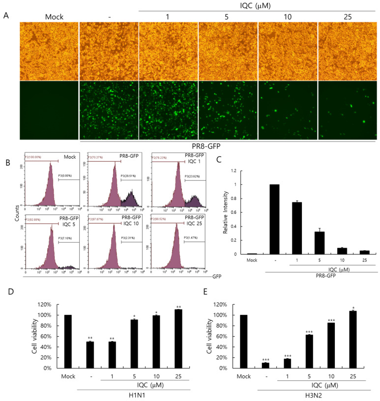
Figure 2.
The antiviral effect of isoquercitrin against influenza A viral infection. (A-C) IQC at 0, 1, 5, 10, or 25 μM with 10 MOI PR8-GFP IAV were mixed for 1 h at 4 °C, the mixtures were cotreated to RAW 264.7 cells for 2 h at 37 °C. After washing with PBS, the cells were further incubated for 24 h at 37 °C. (A) Brightfield and fluorescence images were obtained using the fluorescence microscope with 200× magnification. (B,C) The cells were fixed with 4% paraformaldehyde and analyzed by FACS. The levels of GFP expression were depicted as relative intensities compared to the PR8-GFP IAV-infected control. The data represent the mean ± SD based on three independent experiments. (D) H1N1 or (E) H3N2 IAV at 50 MOI was incubated with IQC at the indicated concentrations 1 h at 4°C and the mixture was added to the cells until cytopathic effect formation at 37 °C. The supernatant was harvested to assess the cell viability using a CCK-8 assay. The data represent the mean ± SD based on three independent experiments. Statistical significance was assessed via an unpaired Student t-test. *** p < 0.001, ** p < 0.005, * p < 0.05.

