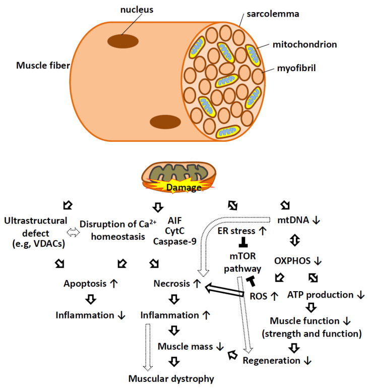
Figure 3.
Schematic representation of mitochondrial dysfunction as causative factors in muscular dystrophy. In skeletal muscle fibers, mitochondria are located between myofibrils, surrounding the nucleus, and at high density within the sarcolemma. Mitochondrial dysfunction can manifest through loss of structural integrity during MOMP and MIMP and result in the release of CYTC, AIF, caspase-9, and calcium into the cytosol, triggering apoptotic and necrotic cell death programs. Ultrastructural defects, insufficient or defective mtDNA replication and aberrant protein expression can lead to impaired oxidative phosphorylation, insufficient respiration, and reduced ATP synthesis. Defects in respiration often result in elevated ROS production that can have consequences beyond the mitochondria. Mitochondrial dysfunction often connects with ER stress in that both can activate various signaling pathways (e.g., MTOR) that interfere with the normal trophic signaling necessary for muscle cell growth and proliferation. Reduced cell replication, increased cell death, and reduced anabolic responses lead to reduced muscle mass in patients with muscular dystrophy. AIF, apoptosis-inducing factor; ATP, adenosine triphosphate; ER, endoplasmic reticulum; mtDNA, mitochondrial DNA; MTOR, mammalian target of rapamycin kinase; OXPHOS, oxidative phosphorylation; ROS, reactive oxygen species; VDAC, voltage-dependent anion channel. Solid arrows indicate direct effects, while the dotted arrow indicates indirect effects. The ↑ bar indicates an increase, while the ↓ bar indicates a decrease. The black T bar indicates an inhibitory effect.

